Appendix G – Protocols for Hospital Consults
Protocols for Hospital Consults
Text from this document (searchable)
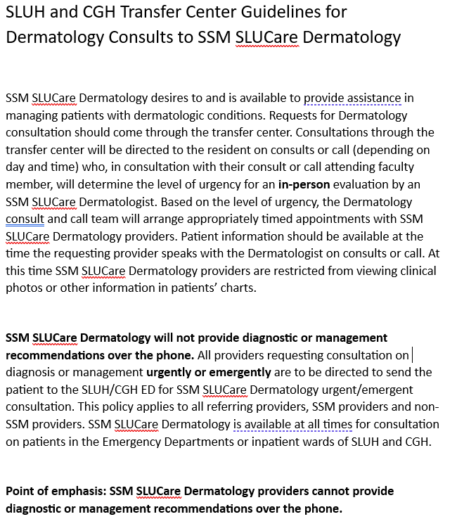
SLUH and CGH Transfer Center Guidelines for
Dermatology Consults to SSM StuCare Dermatology
SSM SLUCare Dermatology desires to and is available to provide assistance in
managing patients with dermatologic conditions. Requests for Dermatology
consultation should come through the transfer center. Consultations through the
transfer center will be directed to the resident on consults or call (depending on
day and time) who, in consultation with their consult or call attending faculty
member, will determine the level of urgency for an in-person evaluation by an
SSM SLUCare Dermatologist. Based on the level of urgency, the Dermatology
consult and call team will arrange appropriately timed appointments with SSM
SLUCare Dermatology providers. Patient information should be available at the
time the requesting provider speaks with the Dermatologist on consults or call. At
this time SSM SLUCare Dermatology providers are restricted from viewing clinical
photos or other information in patients’ charts.
SSM Su Care Dermatology will not provide diagnostic or management
recommendations over the phone. All providers requesting consultation on|
diagnosis or management urgently or emergently are to be directed to send the
patient to the SLUH/CGH ED for SSM SLUCare Dermatology urgent/emergent
consultation. This policy applies to all referring providers, SSM providers and non-
SSM providers. SSM SLUCare Dermatology is available at all times for consultation
on patients in the Emergency Departments or inpatient wards of SLUH and CGH.
Point of emphasis: SSM SLUCare Dermatology providers cannot provide
diagnostic or management recommendations over the phone.
Pager: You are expected to be easily reached if you are on call, please make sure that the call/consult pager is not out of battery! Also make sure that you are accessible by cell phone and/or personal pager when you are on call so that the residents on Consults or attending can easily reach you. If you are paged to a five digit number it means that it is from SLUH and you can call the operator 314-257-8000 to transfer you. If it is a four digit number it is from Cardinal Glennon and you should call the CG operator 314-577-5600 to connect you. If you are paged to the call pager number (419-7186) it means you have a message on your pager, dial the pager number 314-419-7186 then press 0 then the access code 4444 then 3 to hear the message. Same goes for your personal pager, if you are paged to your own number it is because there is a message. Follow the same instructions access code is either 4444 or 1234. Call officially starts at 5 PM; if you are called regarding a consult before then; please have them page the consult (314-419-7187) resident. Remember to log your call hours in New Innovations as well as updating the consult log. On Friday afternoons and afternoons prior to holidays, the consult resident should email the on-call team (including the consult and call attending physicians) with a list of current patients and things that need to be followed up on over the weekend. On Sunday evenings and evenings following a holiday, the call resident should email the consult and on-call teams (including the consult and call attending physicians) with a list of current patients and things that need to be followed up on. If Dr. Siegfried has been involved with any patient care over the weekend or holiday, she also needs to be included on this email. Always remember to use area codes when calling back pages.
General Information:
Consult Notes: Use the Smarttext to generate the Dermatology Consult note in EPIC. If a biopsy is performed, a procedure note must be completed at the end of the consult note—this should not be a separate note. Subsequent day notes should be progress notes—inpatients we are following should be seen every weekday and a note should be placed on the chart until the consult attending agrees that Dermatology can sign off. For phone call related documentation, a telephone encounter or documentation only encounter should be created (Plan of Care notes should not be used as these are nursing notes and not meant for physicians!)
Billing: Every new consult that is staffed will need the diagnosis added to the patient’s problem list so the attending can associate it for billing.
Every consult needs to be added to the Dermatology Consult Handoff template in Epic (see below) as well as the consult log document.
Biopsy for H&E (see biopsy protocol PowerPoint link below): Consult bag should have equipment needed (alcohol, punch, triple pack, suture, dressing supplies). The biopsy bag is to be carried with you so that you have supplies ready at bedside when you get a consult. The consult bag CANNOT be left down in the Central Distribution Room. Again, it is to be carried with you during consult hours and stored in resident room or locker at CSM after hours. It is the consult resident’s responsibility to monitor the bag for supply stores and order refills from central supply. It usually takes approximately 2-3 days for new supplies (ie biopsy kits, suture, etc) to come in. Supply refills will be left in the Central Supply room for the consult resident to pick up. It is the consult resident’s responsibility to pick up supply refills as soon as they come in and restock the biopsy bag. At SLUH you will need to order lidocaine and ask the nurse to retrieve it for you (do this first, time consuming). A NURSE MUST BE PRESENT TO PERFORM A TIME-OUT BEFORE STARTING BIOPSY. Label specimen bottle with patient’s sticker. No need to have secretary put a special order through computer. Fill out pathology specimen sheet at CG (Pathology Request for CG found either in bag or at nurses’ station) OR if at SLUH place the order for “specimen tissue” with your diagnosis and the path form will print out. Take specimen and path sheet (in specimen bag) to the dermatopathology lab. If an after-hours or weekend biopsy, send an Epic message to the Dermpath Pool to alert them that a specimen is needing to be processed.
PATHOLOGY RESIDENT ON CALL: 314-490-1402DERMATOLOGY CONSULT PAGER: 314-419-7187 (M-F, 8AM-5 PM)DERMATOLOGY ON CALL PAGER: 314-419-7186 (all other hours outside of consult hours)
Inpatient Biopsy Protocol.pptx
Skin Biopsy for Bacterial/Fungal/AFB Culture: Write order for bacterial culture, bacterial smear, fungal culture, fungal smear, AFB culture, AFB smear (or whatever is appropriate for that pt), give order sheet to secretary and have her enter into computer. You may also order these yourself and ask the nurse to release the orders. Do this ASAP during the consult because sometime it takes a while to enter and print. Several stickers will print. Put specimen in sterile urine cup with sterile saline (in pink tubes or can use the flushes for IVs), on sterile gauze if desired. Label cup with patient’s sticker. Pull off appropriate lab stickers, date, time, and initial them and put them on cup too. Put specimen and remaining stickers in specimen bag and hand carry to lab. At SLU— on 4th floor at specimen drop off (see above), hand directly to lab worker. At CG— on Ground floor, follow signs to laboratory (in between ER and Radiology), leave at window with lab worker.
Computer orders if the clerk is unsure:
At SLU— go under New Order
CTIStissue bacterial culture with gram stain
CFUN-HSNfungal culture
FUNSfungal smear
CAFBAFB culture
AFBSAFB stain
At CG – go under LAB
A whole list of cultures comes up, search for appropriate (bacterial, fungal, etc) and enter SKIN as source.
Bacterial/Fungal/Viral Swab Cultures: Blue top swab for bacterial, red top for fungal, special shorter red-top with separate swabs for viral. Extra bacterial and fungal culture swabs are in the Utility rooms. If out of viral cultures, can call microbiology laboratory at SLU to have a viral culture (a tiny tube with pink media) tubed to the floor. At Glennon, viral cultures may be available on the floor or can call the laboratory to send one. Write order for the appropriate swab culture and have secretary put through computer to print up stickers. Label specimen, include it with stickers in specimen bag – hand carry to labs as above.
Computer orders if the clerk is unsure:
At SLU— go to orders, enter as:
CULTbacterial culture
GRAMgram stain
CFUN-HSNfungal culture
VCGgeneral viral culture (HSV, VZV, CMV, Cocksackie, Adenovirus)
HERCHSV culture only
At CG – go under LAB
A whole list of cultures comes up, search for appropriate (bacterial, fungal, etc) and enter SKIN as source.
DFA(Direct fluorescent antibody): Use 15 blade to scrape the base of a blister after unroofing (more important to get skin cells than blister fluid). Smear into wells on special slide (in consult/call bag, have yellow film with circular wells). Need to give 2 samples (either 2 wells or 2 separate slides) if want HSV and VZV. Sometimes helpful to use a pen and circle the well/wells that you have put specimen in so lab does not confuse. Place slide in carrier or specimen cup with patient’s label. Write order for DFA, be sure to specify for HSV or VZV or both. Have secretary enter DFA into computer (may need to help her find it) or order it yourself. Take printed stickers and write pager number on labels so they can page you with results. Take to virology lab which is in tunnel between SLU (ground floor) and CG (basement). Glennon does DFA for both hospitals and they usually only run these before 4:30pm. They usually call you with results within an hour.
Computer orders if the clerk is unsure:
At SLU – go under Orders
HERPDFA
VARDFA
At CG – go under LAB
DIR- HSV
DIR- VZV direct fluorescent antibody for VZV and HSV done at Cardinal
Glennon
Tzank: Take 15 blade and unroof blister, scrape base and smear thinly on slide. Stain with Wright stain for about 1 minute then rinse off and lightly pat dry. Usually can take to pathology at 4th floor at SLU or 1st floor at Glennon and ask to use one of their microscopes. They are usually very welcoming if you just introduce yourself and ask to use the microscope for a few minutes.
KOH/Scabies Prep: perform as usual and use same microscope to examine for Tzank.
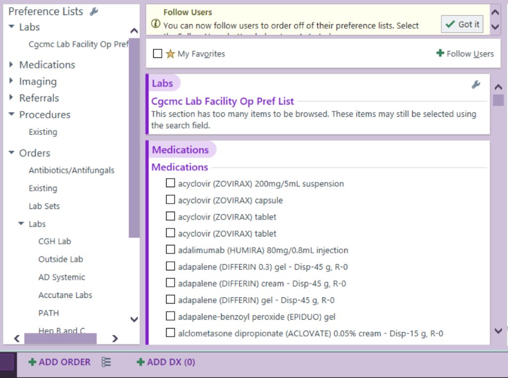
CG Order Sets:
Menu can be accessed by clicking the 3 line icon on the lower left corner, just to the right of “ADD ORDER”; scroll to “Lab Sets” for orders related to CG inpatient skin care and Hemangeol initiation
Text from this document (searchable)
Preference Lists &
- Labs
Cgomc Lab Facility Op Pref
• Medications
• Imaging
• Referrals - Procedures
Existing
• Orders
Antibiotics/Antifungals
Existing
Lab Sets
7 Labs
CGH Lab
Outside Lab
AD Systemic
Accutane Labs
PATH
Hen B and C
- ADD ORDER
Follow Users
• You can now follow users to order off of their preference lists. Select
- 11
V Got it
- My Favorites
- Follow Users
Labs
Cgomc Lab Facility Op Pref List
This section has too many items to be browsed. These items may still be selected using
the search field.
Medications
Medications
LI acyclovir (ZOVIRAX) 200mg/5mL suspension
• acyclovir (ZOVIRAX) capsule
• acyclovir (ZOVIRAX) tablet
L acyclovir (ZOVIRAX) tablet
LI adalimumab (HUMIRA) 80mg/0.8mL injection
• adapalene (DIFFERIN 0.3) gel - Disp-45 g, R-0
LI adapalene (DIFFERIN) cream - Disp-45 g, R-O
• adapalene (DIFFERIN) gel - Disp-45 g, R-O
• adapalene-benzoyl peroxide (EPIDUO) gel
• alclometasone dipropionate (ACLOVATE) 0.05% cream - Disp-15 g, R-O - ADD DX (0)
^
Biopsy request in the OR/procedural suite unit at CGCH
- Refer to Appendix OO
Kawasaki’s Disease
Kawasaki’s disease is a systemic vasculitis involving the small and medium-sized muscular arteries, especially coronary arteries. It occurs predominantly in children under age 5, peaks between 1-2 years, and favors boys. Kawasaki’s disease is a diagnosis of exclusion. Patients who do not fulfill classic criteria are labeled “atypical.” This usually occurs in kids less than 1 year of age (need to maintain a high index of suspicion). Diagnostic criteria includes:
-Fever of at least 5 days duration (initial and cardinal feature)
-Presence of at least four of the following features:
Changes in the extremities (erythema and swelling)
Polymorphous exanthem
Bilateral conjunctival injection
Changes in the lips and oral cavity (strawberry tongue, chelitis)
Cervical LAD of at least 1.5 cm in diameter (least common finding, usually unilateral)
The skin involved in 99% of cases. The first sign usually diffuse erythema and painful induration of hands/feet. A polymorphous exanthem usually appears within 5 days of onset of fever. Perineal macular erythema is characteristic and begins to desquamate within 48 hours. One to three weeks after onset of disease eruption begins to desquamate beneath nail plate and this extends to involve entire palm/sole. Cardiac conditions are the main cause of long-term morbidity/mortality. Coronary aneurysms develop in 20% of untreated patients.
Pathogenesis:
Generalized immune activation with production of various cytokines that cause inflammation and damage to endothelial cells
Differential diagnosis:
Viral infection/exanthem, strep infection, JRA, EM, SSS, TSS
Labs:
Acute phaseà elevated ESR, leukocytosis with left shift, elevated IgG and IgM levels
Subacute phaseà marked thrombocytosis (platelet count up to one million)
Biopsy:
Non-specific
Treatment:
-Baseline and serial cardiac echocardiograms
-IVIG (check IgA level prior to first dose) single dose of 2g/kg over 10-12 hrs
-high-dose aspirin (100 mg/kg/day) divided QID
-High-dose methylprednisolone for treatment failures
-Treatment for atypical cases should be based on clinical judgment
Vesiculopustular eruptions in the neonate/child
Obtain detailed obstetric and neonatal history (see box)
Differential diagnosis:
Infection (Staph aureus, group A Strep, Candida, HSV, scabies)
Transient skin lesions (neonatal pustular melanosis, erythema toxicum neonatorum, cephalic pustulosis)
Rare causes: acropustulosis of infancy, eosinophilic pustular folliculitis, incontinentia pigmenti
Work-up:
Tzanck smear (may need to do multiple sites)
-see multi-nucleated giants cells in viral disease
-take to lab at CGCH and ask for Wright stain
DFA
-scrape base of blister onto slide (test done at CGCH)
-need one slide for HSV/VZV, two slides to type HSV
Viral PCR and culture
-skin scraping of the base of an unroofed vesicle
-obtain with a #15 blade
-place immediately on ice
-if HSV suspected then culture conjunctiva, throat, CSF, urine
Bacterial smear and culture (culturette swab sample)
KOH
-positive in candidiasis
Gram stain
-useful to identify transient skin lesions (see eosinophils in erythema toxicum neonatorum, neutrophils in transient neonatal pustular melanosis)
Biopsy for H&E
Biopsy for bacterial and fungal culture (needs to be placed in sterile saline)
Treatment
Will be guided by work-up
If high suspicion of HSV, then isolate infant and initiate empiric therapy promptly (acyclovir 10 mg/kg IV q8h for 10 days)
Bullous eruptions in the neonate/child
Differential diagnosis:
Infections (SSSS, GBS, Pseudomonas, congenital syphilis, fetal varicella, intrauterine HSV)
Transient lesions (sucking blister, birth trauma)
Solitary/few grouped bullae on lower extremities bullous arthropod reaction m/c
Uncommon causes (EB, EHK, mastocytosis, maternal bullous d/o)
-Consider linear IgA disease (chronic bullous disease of
childhood) or other primary immunobullous disease in non-neonates
-DIF on perilesional skin will show linear IgA deposition
along DEJ
Work-up:
Tzanck smear (may need to do multiple sites)
DFA
Viral PCR and cultures of skin
Bacterial culture of skin
KOH
Gram stain
Biopsy for H&E
Biopsy for bacterial and fungal culture (needs to be placed in sterile saline)
Biopsy of perilesional skin for DIF
-useful for primary bullous disease
Treatment
Will be guided by work-up
‘
Eruptions in Immunocompromised Patients
Includes heme/onc, transplant, HIV/AIDS patients
BE AGGRESSIVE!!! These patients can decompensate rapidly and the clinical presentation can be varied
| PREDISPOSITION | INFECTION |
| Neutropenia/functional neutrophil defect | Aspergillosis, oropharyngeal and/or systemic candidiasis, zygomycosis, infections due to rare organisms |
| CD4 lymphopenia (AIDS) | Oropharyngeal candidiasis, cryptococcosis and endemic respiratory mycoses such as histoplasmosis, nocardiosis |
| Diabetes mellitus | Zygomycosis |
| Heart valve surgery | Various but mainly C. albicans and non- albicans Candida spp |
General work-up for suspected deep fungal infection/candidiasis
-Biopsy for H&E
-Biopsy for tissue culture (sample should be large enough for fungal,
bacterial, and mycobacterial- no smaller than 3 mm punch)
-KOH of lesions
-Gram stain, AFB smear, and KOH of tissue can be done quickly and
can aid in treatment while awaiting culture results
-fine-branching filaments on Gram stainàNocardia
-Septated, acute-branching fungiàAspergillus
-Pseudohyphae and yeast formsà Candida
General work-up for suspected viral infection
-Tzanck prep, viral culture, and DFA
-Biopsy for H&E
-Ophthalmology consult if necessary
-Isolation
-Institute IV anti-viral therapy promptly
Acute disseminated histoplasmosis
-occurs in untreated AIDS patients (skin lesions are manifestation of disseminated infection to liver, spleen, bone marrow, and lymphoreticular system)
-present with fever and progressive weight loss
-clinically may see papules, small nodules, molluscum-like lesions that may develop into shallow ulcers
-diagnosis confirmed with biopsy and culture (H. capsulatum is an intracellular parasite often seen in macrophages)
-serologic tests for detection of circulating Histoplasma antigen available
-treatment: depends on severity of disease
itraconazole 200-400 mg daily or IV amphotericin B (up to 1 mg/kg/day)
Cryptococcosis
-most common clinical manifestation is meningoencephalitis
-acneiform papules or pustules progressing to warty or vegetating, crusted plaques, ulcers and hard infiltrated plaques or nodules characteristic of widespread systemic infection
-diagnosis confirmed with biopsy and culture (capsules of cells can be stained with mucicarmine or Alcian blue stains; not difficult to organism to grow in culture)
-treatment with IV amphotericin B and flucytosine in non-AIDS patient (add long-term fluconazole in AIDS patients due to high relapse rate)
-in patients with few skin lesions only, can use fluconazole or itraconazole
Aspergillosis
-clinically can see large necrotic lesions such as ecthymagangrenosum; also smaller papules and cold abscesses
-treatment with amphotericin B
Fusarium
-widely distributed target-like lesions that may undergo central necrosis; digital cellulites and superficial white onychomycosis
-treatment with amphotericin B
Systemic candidiasis
-skin eruption follows dissemination from GI tract or via bloodstream
-neutropenic patientsà severe disseminated disease with widespread skin nodules and associated muscle pains
-treatment with IV amphotericin B or fluconazole
HSV
-same diseases as those seen in immunocompetent hosts but more severe, extensive, and difficult to treat
-patients with defects of T-cell immunity at particular risk for progressive mucocutaneous or visceral infections (degree of dissemination depends on level of host immunodeficiency)
-recurrent and persistent ulcerative HSV lesions very common among patients with AIDS
-HSV may involve oropharynx, esophagus, tracheobronchitis, and pneumonitis
-treatment for disseminated disease is IV acyclovir 5-10 mg/kg q 8 hours given until vesicular lesions begin to crust over (15 mg/kg q 8 hours for life-threatening disease)
Varicella
-continued virus replication and dissemination result in a prolonged high-level viremia, a more extensive rash, and a longer period of new vesicle formation
-pneumonia, hepatitis, encephalitis, and hemorrhagic complications may develop
-can range from mild febrile purpura to severe and often fatal purpurafulminans/ “malignant” varicella
Herpes zoster
**-**vesicles are present on the nasal tip (Hutchinson’s sign)à this represents trigeminal nerve involvement
-ophthalmology consult
GVHD
-m/c cause of morbidity and mortality in allogeneic stem cell and
BMT pts
-classically divided into acute (<100 days post-tx) and chronic (>100
posttx)
-Acute GVHD
-major sites of involvement are skin, liver (hepatitis), GI tract (diarrhea)
-morbilliform eruption (grading system for %BSA stages I-IV)
-mucosal surfaces can be involved
-early GVHD athology can be non-specific (lichenoid)
-check CBC, LFTs
Staphylococcal Scalded Skin Syndrome (SSSS)
-acute life-threatening disease of infants/toddlers caused by exotoxin produced by Staph aureus (group II phage 71 m/c)
-exotoxin targets desmoglein-1
-adults with renal compromise or immunosuppresion predisposed
-begins with sudden onset of fever and tender blanchable erythema; begins peri-orally and spreads quickly to remainder of body, flaccid blistering occurs within 24-48 hours, with subsequent exfoliation in large sheets (+ Nikolsky’s sign)
-palms, soles, mucus membranes spared
Differential diagnosis:
-TEN (see full-thickness epidermal necrosis on path, do frozen section for rapid interpretation)
-Diffuse cutaneous mastocytosis
-Exfoliative erythroderma
-Sunburn
-Thermal burn
-TSS
-Streptococcal scarlet fever
-Kawasaki’s disease
Diagnosis:
-jelly roll sample of desquamated skin for urgent frozen section (to rapidly distinguish from TEN)
-Biopsy see split through granular layer on H&E
-Cannot culture Staph from bullae because lesions are caused by exotoxin
-may recover Staph from blood, urine, pharynx, conjunctiva
Treatment
-No steroids!!!
-Broad-spectrum penicillin
-Electrolyte monitoring
-Wound care
- Eczema Herpeticum (Kaposi’s Varicelliform Eruption)
1) History/physical exam/pertinent positives/negatives:
-
A distinct cutaneous eruption caused by herpes simplex virus type 1 (HSV-1), herpes simplex virus type 2 (HSV-2), coxsackievirus A16, or vaccinia virus that infects a preexisting dermatosis (atopic dermatitis, seborrheic dermatitis, scabies, Darier’s, Hailey-Hailey, pemphigus). Most commonly, it is caused by a disseminated HSV infection in patients with atopic dermatitis.
-
clusters of monomorphic umbilicatedvesiculopustules in areas where the skin has been affected by a preexistent dermatitis, most commonly on head & neck.
-
Umbilicatedvesiculopustules that progress to punched-out erosions that can be hemorrhagic and crusted in the setting of a widespread dermatosis is virtually pathognomonic for Kaposi varicelliform eruption. Can have the characteristic “buckshot”appearance.
-
Associated with a fever, malaise, and regional lymphadenopathy.
-
These erosions may coalesce to form large, denuded areas that frequently bleed and can become secondarily infected with bacteria.
- Differential Diagnosis
-
Chicken Pox (Varicella Zoster)
-
Impetigo/ Bullous Impetigo/ Impetiginized eczema
- Patient work-up
-
DFA staining of scrapings from the base of an early vesicular or crusted lesion is as accurate as viral culture in differentiating HSV-1, HSV-2, and varicella-zoster virus (VZV).
-
PCR and viral culture placed immediately on ice
At CGH: can enter all orders and have nursing verify to print out sticker then these specimens can be taken to the SSM virology lab (offsite, at SJW Hospital); They’re only open Mon-Fri 8-4:30 pm and variably Sat am so if after hours can call the lab. You need to tell them to run it for HSV or VZV and if both they need two slides
At SLUH: Bring to the fourth floor lab window. Reminder: This is a send out lab so will take several days to result.
-
A Tzanck smear of the base of an opened vesicle or erosion can provide rapid diagnosis when it shows the characteristic epithelial multinucleated giant cells and acantholysis.
-
Skin biopsy if diagnosis is uncertain to rule out other etiologies.
-
Ophthalmologic consultation is indicated for scleral injection, photophobia an/or nasociliary nerve (nasal tip) involvement. Herpetic keratitis can lead to scarring.
- Treatment
-
Discontinue topical steroids
-
IV Acyclovir 10 mg/kg/d divided tid for 5 d or until lesions crust over, or valacyclovir (10 mg/kg BID-TID for 10-14 d or until lesions heal. Liquid formulation valacyclovir is only available if compounded. Use with caution in renal failure (check BUN/Cr)
-
For children, Acyclovir 20 mg/kg/dose TID x 5 days or Valacyclovir po
-
Cover with anti-staphlyococcal anti-biotics for secondary bacterial co-infection.
- Purpuric eruptions including purpura fulminans
1) History/physical exam/pertinent positives/negatives:
- Family history of bleeding or thrombotic disorders, duration of symptoms, drugs and medications that might affect platelet function/coagulation (warfarin use, Heparin inHIT), history of liver disease/alcoholism/hepatitis, collagen/vascular dz, APA., recent procedures (cardiac cath), recent trauma, recent viral illnesses, neurologic symptoms (HA, confusion), asplenic (think meningococcemia), epistaxis, immune status (gram neg sepsis in immunocompromised)
- Exam documenting the size, type (palpable/nonpalpable), distribution of purpura (typically dependant legs and ankles), any telangiectasias, feel for splenomegaly/hepatomegaly, look for signs of liver disease, underlying malignancy, any joint problems, presence of fever, lymphadenopathy, vitals c/w shock (DIC), Raynaud’s
- Differential Diagnosis
| Non-Palpable Purpura | Palpable purpura (LCV=small vessels) |
| Coagulopathies – DIC, liver dz, Protein C/ S def, APA | Idiopathic - #1 cause |
| Thrombocytopenia - ITP/TTP, BM suppression | Drug (ASA, NSAIDs, sulfa, PCN, thiazides) |
| Thrombocythemia – plt>1,000,000 | Infection (strep, RMSF, GC, meningiococ, TB, Hepatitis, other viruses, syphilis |
| Drug – anticoag (warfarin nec/HIT), steroids, NSAIDS | |
| Infection – SBE, RMSF, meningococc, GC, TORCH | Collagen Vascular (livedoid) – RA, SLE, DM |
| Scurvy | Carcinoma – lymphoma, leukemia, mult myelo |
| Senile purpura (increased skin fragility) | Cryos – cold, mixed, mm, hep, CA, idiopathic |
| Thrombotic/embolic – cholest emboli, marantic, fungi | Cold agglutins – viral pneumonia, SLE, lymphoma |
| Livedoreticularis | HSP |
| Traumatic | Med vessel – PAN, Wegeners, Churg-Strauss |
| Systemic disease – EDS, amyloidosis | |
| Capillaritis (Schambergs, majocchi’s, etc.) | Livedoidvasculitis (atrophie blanche) |
| Bland Thrombi (cryoglobulins, fibrin) | Sweet’s syndrome |
- Patient work-up
-
CBC,UA, LFTs, Hep B&C, BldCx, ANA, BUN/Cr, ESR, SPEP, PT/PTT/INR, fibrin, fibrinogen, fibrin split products, P-ANCA, C-ANCA, ASO-titer, serum cryoglobulins, echo
-
Gram stain of pustules/purpura to r/o infectious cause of purpurafuluminans
-
Skin biopsy for H & E (LCV shows fibrinoid necrosis of blood vessel wall and fragmentation of nuclei, Purpurafulminans), consider DIF
- Treatment
-
Supportive care (rest and elevation of affected extremity, eliminate identified drug)
-
Consider IV Heparin, Vitamin K or FFP transfusions if appropriate for coagulopathy
-
NSAIDS/Antihistamines for vasculitis confined to the skin
-
Consider systemic steroids(60-80 mg/d) if vasculitis with systemic manifestations or necrotic lesions
8a. TEN/SJS
1) History/physical exam/pertinent positives/negatives:
-
Constitutional symptoms, such as fever, cough, or sore throat, may appear 1-3 days prior to any cutaneous lesions and may be a sign of causative bacterial or viral infection (mycoplasma & HSV assoc with EM/SJS).
-
HIV positive patients at 1000 fold increased risk
-
Drug exposure timeline is essential, typically 1-3 weeks between drug exposure & rash.
-
Drugs most commonly implicated include: trimethoprim-sulfamethoxazole and other sulfonamide antibiotics, aminopenicillins, quinolones, and cephalosporins, anticonvulsants, NSAIDs, allopurinol,and barbiturates.
-
Patients may complain of a burning sensation in their eyes, photophobia, painful urination/ defecation, difficulty breathing. Recent cold sores/history of HSV.
-
Initial skin lesions are macular and desquamate, form atypical targets with purpuric centers that coalesce, form bullae then slough.
-
Nondenuded areas have a wrinkled paper appearance.
-
Positive Nikolsky sign is easily demonstrated by applying lateral pressure to bullae.
-
More than 2 mucous membranes are involved. Painful oral erosions cause severe crusting of the lips, increased salivation, and impaired alimentation.
-
A recent classification proposes that epidermal detachment in SJS is less than 10% of BSA. 10-30% of BSA is TEN/SJS overlap. TEN has detachment of more than 30%. Accurate BSA estimate of involvement is crucial.
- Differential Diagnosis
-
EM Minor- Bullous Drug reaction
-
Staph Scalded Skin Syndrome- Morbillifom drug eruption
-
BP, PV, Paraneoplastic Pemphigus- Drug-Induced Pemphigus
-
Acute GVHD- Chemical Burn
-
Pustular Psoriasis- Bullous Lupus
-
Linear IgA Dermatosis- AGEP
- Patient work-up
- Skin Biopsy for H&E (can do frozen section/jelly roll prep to differentiate from SSS for stat evaluation of level of split and if full-thickness necrosis), DIF if can’t r/o blistering d/o
-CBC, BMP, LFTs, PT/PTT, bacterial cultures of blood and areas of denuded skin, serum IgA level in anticipation of IVIG therapy, CXR
- Ophtho consult to evaluate for ocular involvement, consider GI/Pulm/Urology consult
- Treatment
-
Withdrawal of causative meds
-
Steroids are controversial as increase mortality but may be of value early in high doses to stop the evolution, then quickly withdrawn to prevent increased risk of sepsis.
-
IVIG 1gm/kg qd x 3 days for total 3mg/kg dose – the earlier the better the prognosis
-Consider TNF-alpha inhibitor
- Treat like burn patient (fluids/wound care/ nutrition/sterile technique/transfer to ICU/burn unit )
8b. DRESS (Drug Reaction with Eosinophila and Systemic Symptoms)
1) History/physical exam/pertinent positives/negatives:
-
Fever and rash are most common symptoms
-
Drug exposure timeline is essential, typically 2-6 weeks after drug exposure (later).
-
Drugs most commonly implicated include: aromatic anticonvulsants (phenytoin, phenobarbital, carbamazapine) sulfonamides, dapsone, minocycline, allopurinol
-Brown recluse spider envenomation (loxocelism)
-
Starts as morbilliform eruption, becomes edematous with follicular accentuation on face, trunk and UE. Can develop vesicles and tense bullae. Edema of the face is classic.
-
Lymph nodes enlarged, arthralgias, arthritis
-
Hepatitis (10% fatal), myocarditis, interstitial pneumonitis/nephritis, thyroiditis seen.
-
Peripheral eosinophilia is common and characteristic.
- Differential Diagnosis
- Morbillifom drug eruption
- Bullous Drug reaction
- Viral syndrome
- Hypereosinophilic synd
- Lymphoma
- Pseudolymphoma
- Edema Blisters
- Patient work-up
- Skin Biopsy for H&E
- CBC with diff looking for eosinophilia, BMP, LFTs, CXR, EKG
- Treatment
-
Withdrawal of causative meds
-
Corticosteroids are first line for DRESS with slow taper to avoid relapse
-
Topical steroids may help symptomatically in mild cases
8c. Reactive Infectious Mucocutaneous Eruption (RIME)
Clinical Presentation:
-
Mucositis affecting 1 or more mucosal sites including oral, conjunctival, urogenital mucosae (less commonly esophageal/anal involvement) with or without cutaneous involvement
-
Cutaneous involvement consists of typical or atypical targetoid lesions (two rings) +/- bullae or vesicles affecting any area of the body; acral surfaces commonly affected
-
Often triggered by infections; symptoms commonly preceded by cold/flu symptoms by approx 1 week +/- fever ≥100.4°F for ≥24 hours
-
Most common causative agent is mycoplasma pneumonia. Others include Chlamydia pneumoniae, rhinovirus, adenovirus, enterovirus, human metapneumovirus, parainfluenza, influenza B virus and COVID 19 Diagnosis: RIME is a clinical diagnosis supported by history and typical mucocutaneous findings in the context of congruent infectious workup and absence of a history of medications known to cause SJS/TEN. Other dx in the ddx include:
-
SJS/TEN ~ hx of a culprit medication
-
EM Major ~ hx of HSV or other common trigger
-
Multifocal fixed drug (+/- bullous) ~ hx of a culprit medication
Lab Work-Up:
- Mycoplasma PCR or Antibodies
- Viral Respiratory Panel—includes the following (Note: 20% false negative rate and no EBV or Parvovirus included)
- Adenovirus PCR
- Coronavirus 229E PCR
- Coronavirus HKU1 PCR
- Coronavirus NL63 PCR
- Coronavirus OC43 PCR
- COVID-19 PCR
- Human Metapneumovirus PCR
- Human Rhinovirus/Enterovirus PCR
- Influenza A PCR
- Influenza B PCR
- Parainfluenza Virus 1 PCR
- Parainfluenza Virus 2 PCR
- Parainfluenza Virus 3 PCR
- Parainfluenza Virus 4 PCR
- Respiratory Syncytial Virus PCR
- Bordetella parapertussis PCR
- Bordetella pertussis PCR
- Chlamydia pneumoniae PCR
- Mycoplasma pneumoniae PCR
- Consider COVID specific PCR given high false (-) rate of VRP
- Chest X-ray can support findings of pneumonia in mycoplasma infx
- HSV PCR (oral mucosae); assess for EM triggers
- EBV serologies; assess for alternate RIME/EM triggers
- Biopsy for H&E (taken from non-eroded targetoid lesion) + DIF (perilesional skin)—often biopsy is not necessary especially in children
- IgA level if anticipating tx with IVIG; risk for anaphylaxis with IVIG if IgA deficient
Management:
- Have primary team order consult from pertinent services; Ophtho, Urology, Gynecology, ID
- There is limited data comparing systemic treatment options. Given its safety profile and efficacy for SJS/TEN/EM, IVIG is the preferred systemic tx dosed at 1g/kg/day x3 days.
- If pt symptoms continue to *progress or pt has contraindications to IVIG, second line therapy is Cyclosporine at 3-5mg/kg/day divided into two doses for 7-14 days. *Progression is indicated by the formation of new skin or mucosal lesions; if individual lesions form bullae or slough this is in line with the natural progression of the disease.
- Systemic steroids, etanercept, other TNF-a inhibitors and plasmapheresis are additional alternative systemic treatment options; however, at this time we are unable to get biologic medications as inpatient
- If mycoplasma pneumonia infx is identified, appropriate abx should be initiated; Azithromycin is the most common first line agent; other agents (antiviral vs antibacterials) may be utilized when appropriate depending on the causative agent
- Rash may be treated with high-potency topical corticosteroids on the torso & extremities and low-potency steroids for lesions on the face, genital region, or skin folds
- Lesions of the oral mucosae can be managed with magic mouthwash, cyclosporine rinses, or topical steroid gels.
- Cyclosporine solution is in the EMR as “Cyclosporine microemulsion (Neoral) 100mg/ml oral solution”
- Pt should swish 2-5ml of the cyclosporine solution for 3-5min twice daily
- Pts report that cyclosporine rinses can burn, recommend using rinse immediately following magic mouth wash
- Insurance coverage for cyclosporine mouthwash varies by carrier and the medication is readily available at SLU/CG outpatient pharmacy. St. Peters pharmacy and other specialty pharmacies also carry the medication. Cyclosporine solution has been difficult to obtain from Walgreens/CVS
- Vaseline should be applied regularly to areas of denuded skin as well as non-adherent dressings
Other:
Patients and families should be educated on the expected course of the disease including:
- Possibility that lesions may develop vesicles/bullae as part of the normal disease process (though this does not always occur in RIME)
- Significant shedding of skin in the oral and oropharyngeal mucosae
- Set the expectation that it takes several weeks and up to 1-2 months for the oral mucosae and skin to reconstitute (long recovery time)
- Recurrence rate of RIME is estimated to be anywhere from 8% to 30% so pts should notify their PCP right away if they notice similar symptoms developing in the future
- Pustular/Erythrodermic Psoriasis
1) History/physical exam/pertinent positives/negatives:
-
History of plaque psoriasis and strong family history of psoriasis is common
-
Patients experience constitutional signs and symptoms, such as headache, fever, chills, arthralgia, malaise, anorexia, and nausea.
-
Develop sudden onset pustules and lakes of pus periungually, on the palms, and at the edge of psoriatic plaques ® generalized erythema and more pustules which dry up to form crusts over brown shiny surface. Pustules may occur on the tongue and subungually, resulting in dysphagia and nail shedding, respectively. These pustules coalesce within 1 day to form lakes of pus that dry and desquamate in sheets, leaving behind a smooth erythematous surface on which new crops of pustules may appear. These episodes of pustulation may occur for days to weeks, causing severe discomfort.
-
The patient appears frightened, tachypneic, tachycardic, and febrile. The oropharyngeal mucosa may be hyperemic with geographic tongue. The skin shows a generalized or patchy erythema studded with interfollicular pustules that may have an annular or nonspecific configuration. Flexural and anogenital accentuation may be present. The lesions may appear on the trunk, extremities, and rarely, on the face. Scaling may be observed, especially in areas that already have undergone pustulation.
-
Triggers for flare: Withdrawal of systemic steroids, Drugs (B-blockers, salicylates, iodine, lithium, phenylbutazone, oxyphenbutazone, trazodone, penicillin, hydroxychloroquine, calcipotriol, interferon-alpha, and recombinant interferon-beta injection), irritating topicals (including tar, anthralin, steroids under occlusion, and zinc pyrithione in shampoo), infections, sunlight or phototherapy, cholestatic jaundice, hypocalcemia, Idiopathic in many patients
- Differential Diagnosis
-
SubcornealPustularDermatosis
-
AGEP (drug eruption)
-
Gram-negative septicemia
-
Infected generalized atopic and/or seborrheic dermatitis
- Patient work-up
-
CBC, ESR, Chemistry, UA, blood cultures
-
Tzanck of pustule
-
Culture (bacterial & viral) of pustule
-
Skin Biopsy (psoriasis with neuts/spongiform pustule of Kogoj in upper epidermis)
- Treatment
-
Admission to the hospital to ensure adequate hydration, bed rest, thermoregulation to prevent cardiovascular collapse.
-
Treatment with bland topical compresses, potent topical steroids after saline or oatmeal baths assists in soothing and debriding affected areas. This topical strategy is effective in many pediatric patients as the sole therapy.
-
Initiate systemic therapy with oral retinoids (Soriatane is drug of choice), cyclosporine if flared, methotrexate, or PUVA. If on steroids, taper with caution.
- Cellulitis/Erysipelas/Necrotizing Fasciitis
1) History/physical exam/pertinent positives/negatives:
-
Patients report local pain and swelling at the site of cellulitis.
-
History of Diabetes (predispose to Necrotizing fasciitis), chronic LE edema (elephantiasis nostra from chronic ecurrent cellulitis/erysipelas), tineapedis, recent surgery, skin trauma (eg, scratch, abrasion, animal bites, intravenous drug injection)
-
Fever, chills, malaise may be present
-
Sites are red, hot, & tender with regional lymphadenopathy, streaks of lymphangitis.
-
Unlike erysipelas, borders are poorly demarcated in cellulitis (should mark them daily).
-
Erysipelas classically has raised, well demarcated border because of more superficial dermal involvement, most commonly involving the leg, then face. Fisures, abrasions, leg ulcers may develop gangrene/sepsis
-
Necrotizing fasciitis quickly progresses with dusky blue discoloration, blisters, crepitis, foul odor, anesthesia as it spreads deep to involve fascia, especially the groin (Fournier’s)
- Differential Diagnosis- Erysipeloid (of Rosenbach)
-
Human Bites
-
Impetigo
-
Necrotizing Fasciitis
-
Venous Stasis Dermatitis
-
Deep Fungal
-
Contact dermatitis
-
Eosinophillic cellulitis (Wells syndrome)
-
Sweet’s syndrome
-
Cutaneous mets
- Patient work-up
-
CBC with diff to evaluate for leukocytosis
-
Blood and wound cultures (aerobic and anaerobic) and sensitivities
-
MRI to evaluate for underlying soft tissue involvement if suspect necrotizing fasciitis
-
Skin biopsy if suspect other etiology like contact or lack of response to treatment
- Treatment
-
Mild cellulitis may be treated as outpatient if appropriate oral antibiotics agents with activity against staphylococci and streptococci (eg, dicloxacillin, cephalexin, cefuroxime axetil, erythromycin, clindamycin) are given.
-
More severe disease should be treated initially with intravenous antibiotics in the hospital.Usually cellulitis is presumed to be due to staphylococci or streptococci and is treated with antibiotics (eg, nafcillin, cefazolin). Other options in allergic patients include clindamycin or vancomycin. Ceftriaxone may be useful in the outpatient setting because it can be administered once daily.
-
Consult infectious disease for recommendations on appropriate antibiotic therapy
-
Elevation of involved extremity, support stockings to prevent recurrence
-
If recurrent cellulitis suspected to be due to tineapedis, topical or systemic antifungals
-
Early surgical debridement of necrotizing fasciitis along with polymicrobial coverage
-
If not responding to oral Abx, consider resistance or fungal etiology (crypto, blasto)
PHACES Syndrome
See useful dot phrases:
.PDPHACEIMAGING:
To order MRI/MRA for facial hemangioma patients:
-
Orbit MRI with and without contrast
-
Brain MRI with and without contrast
-
Brain MRA with and without contrast
-
Neck MRA with contrast only
Also, minimum of 50 week post-conception (full-term infants only) eligible for sedation. Sedation order CON124.
Sedation nurse scheduler Mary 268-2700 ext 4205.
-
order test
-
order sedation
-
complete info /tab REF157
Sedation requires NPO x 4 hours if breast fed, 6 hours if formula fed
.PDPROPRANOLOLINPT
HEMANGEOL oral solution contains the beta-adrenergic blocker propranolol hydrochloride and is indicated for the treatment of proliferating infantile hemangioma requiring systemic therapy.
Indications for inpatient initiation: <45 wk gestational age; PHACE syndrome; need for more rapid dose escalation (ulceration, rapid growth, high-risk site, extracutaneous complications (airway involvement, impact on vision)
Guidelines for inpatient initiation
• The recommended starting dose of HEMANGEOL is 0.15 mL/kg (0.6 mg/kg) twice daily, taken at least 9 hours apart.
• Check heart rate, blood pressure, temperature and fingerstick blood sugar 2 hours after each of the first 2 HEMANGEOL doses.
• If heart rate, blood pressure, temperature and fingerstick blood sugar are normal after 2 doses, increase the daily dose to 0.3 mL/kg (1.1 mg/kg) twice daily.
• Check heart rate, blood pressure and fingerstick blood sugar 2 hours after the second 0.3 mL/kg dose.
• If heart rate, blood pressure, temperature and fingerstick blood sugar are normal after 2 more doses, increase the daily dose to 0.4 mL/kg (1.7 mg/kg) twice daily.
To reduce the risk of hypoglycemia, administer HEMANGEOL orally during or right after a feeding. Skip the dose if the child is not eating or is vomiting.
HEMANGEOL is supplied with an oral dosing syringe for administration. Administration directly into the child’s mouth is recommended. Nevertheless, if necessary, the product may be diluted in a small quantity of milk or fruit juice, given in a baby’s bottle.
Hold the dose if:
If Mean Arterial Pressure <40
OR
If heart rate <140 (patient agitated) or <110 (patient at rest) for pt up to 3 months of age
If glucose is <70 (give 30cc of 5% glucose PO, and recheck after 30 minutes).
Collodion Baby
See dot phrase .PICOLLODION
Harlequin Ichthyosis
See Glick JB, Craiglow BG, Choate KA, Kato H, Fleming RE, Siegfried E, Glick SA. Improved Management of Harlequin Ichthyosis With Advances in Neonatal Intensive Care. Pediatrics. 2017 Jan;139(1):e20161003.
Epidermolysis Bullosa
See dot phrase .PIEPIDERMOLYSISBULLOSA
Skin Biopsy for Alpha-Synucleinopathy and Amyloid Inclusion Testing (CND Life Sciences)
Dermatology may be consulted by the neurology inpatient team to perform skin punch biopsies for alpha-synucleinopathy and amyloid inclusion testing through CND Life Sciences. This is a specialized send-out test used in the diagnostic workup of conditions such as Parkinson disease, dementia with Lewy bodies, multiple system atrophy, and pure autonomic failure. The dermatology resident’s role is limited to performing the punch biopsies. Neurology is responsible for consent, requisition paperwork, orders, and specimen submission.
Pre-procedure responsibilities (neurology)
Before proceeding with the biopsies, the dermatology resident should confirm with the neurology team that the following have been completed:
– Informed consent has been obtained from the patient.
– All required CND Life Sciences requisition paperwork has been filled out.
– Necessary orders have been placed.
– The CND Life Sciences biopsy kit is present in the room. Do not use standard dermatology punch biopsy supplies; all materials must come from the CND kit, including the pre-labeled fixative vials containing Zamboni paraformaldehyde solution.
Do not proceed until the kit, consent, and paperwork have been confirmed.
Procedure
The CND protocol requires three 3 mm punch biopsies at the following anatomical sites, taken to the depth of the subcutaneous fat. Always verify the exact site specifications against the instructions included in the current CND kit, as these may be updated over time. Per the published protocol and CND’s standard instructions, the three sites are:
-
Posterior cervical region – 3 cm lateral to the C7 spinous process
-
Distal thigh – 10 cm above the lateral aspect of the knee (lateral femoral condyle)
-
Distal leg – 10 cm above the lateral malleolus
Each specimen must be placed into the specific pre-labeled fixative vial provided in the CND kit. Follow all specimen handling and fixation instructions included in the kit exactly.
Post-procedure responsibilities (neurology)
Immediately after the biopsies are obtained:
– Hand off all labeled specimens and the completed kit directly to the neurology team.
– Do not send specimens through dermatopathology. These are send-out tests processed entirely by CND Life Sciences and managed by the neurology team.
– Neurology is responsible for packaging and shipping the specimens to CND Life Sciences per the kit instructions.