Weedon Outlined
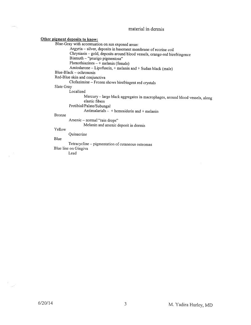
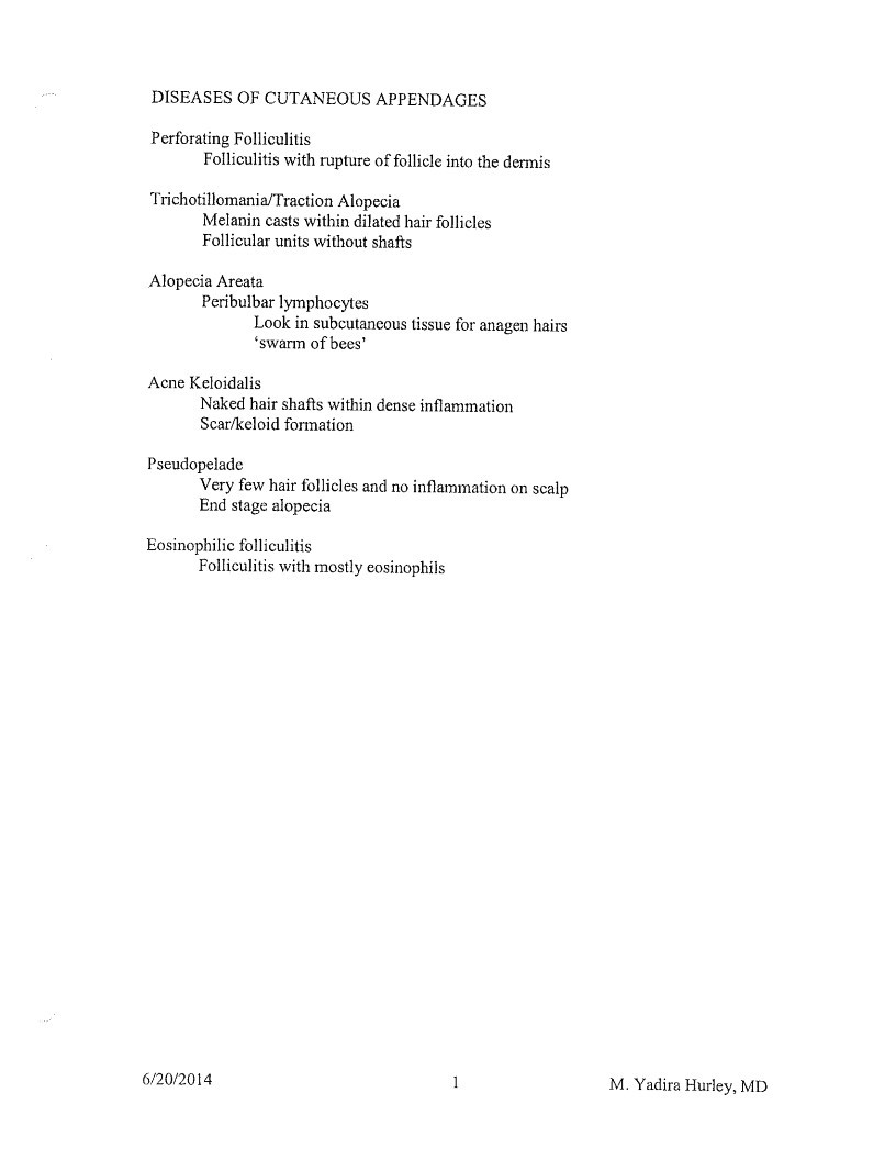
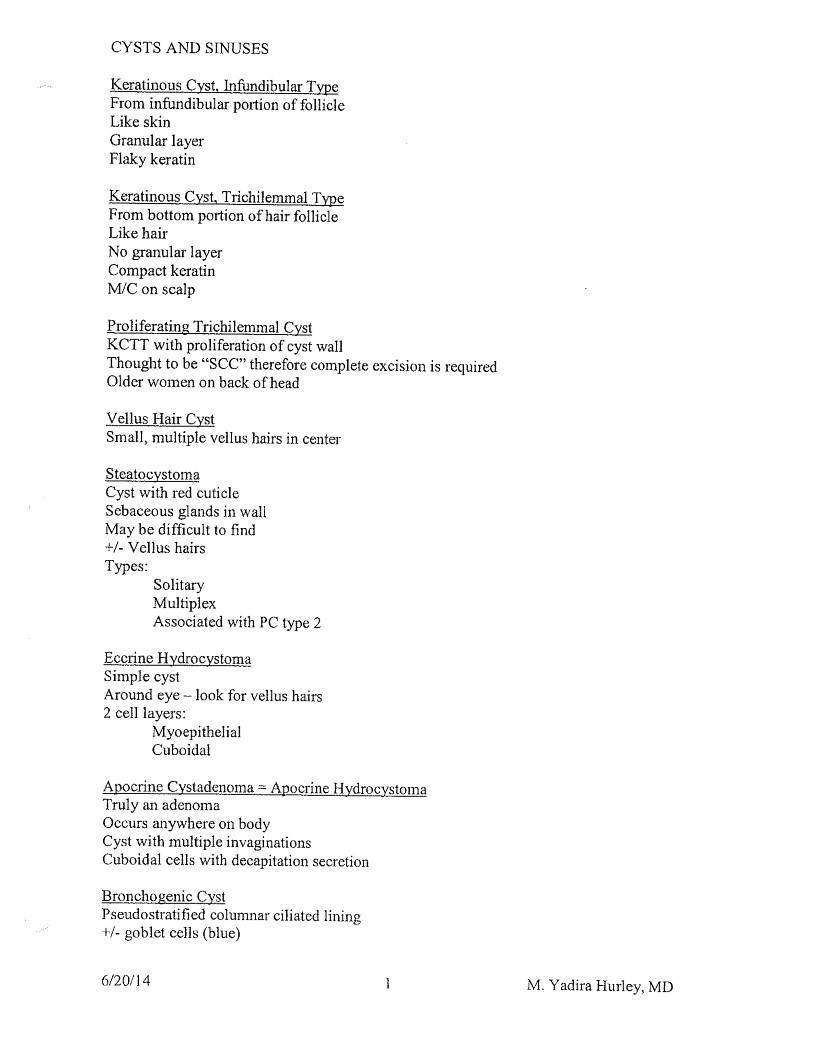
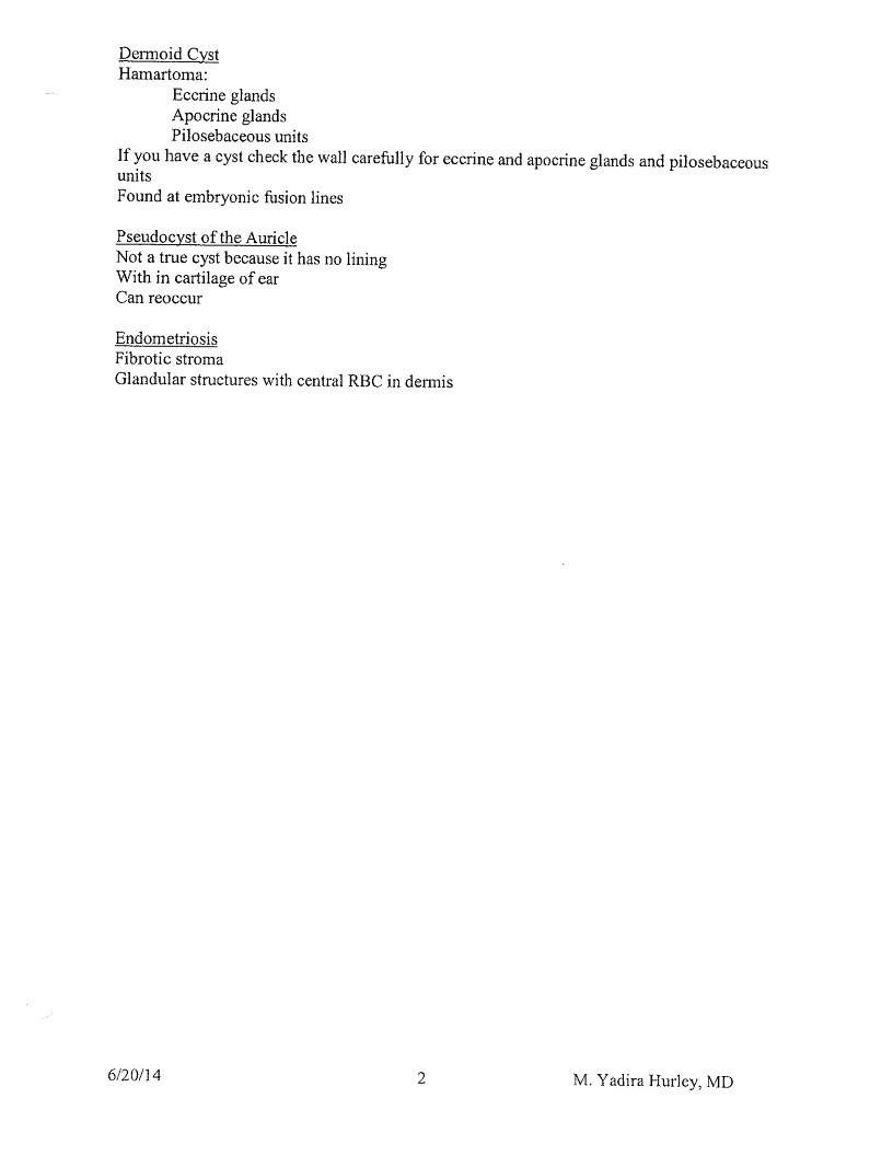
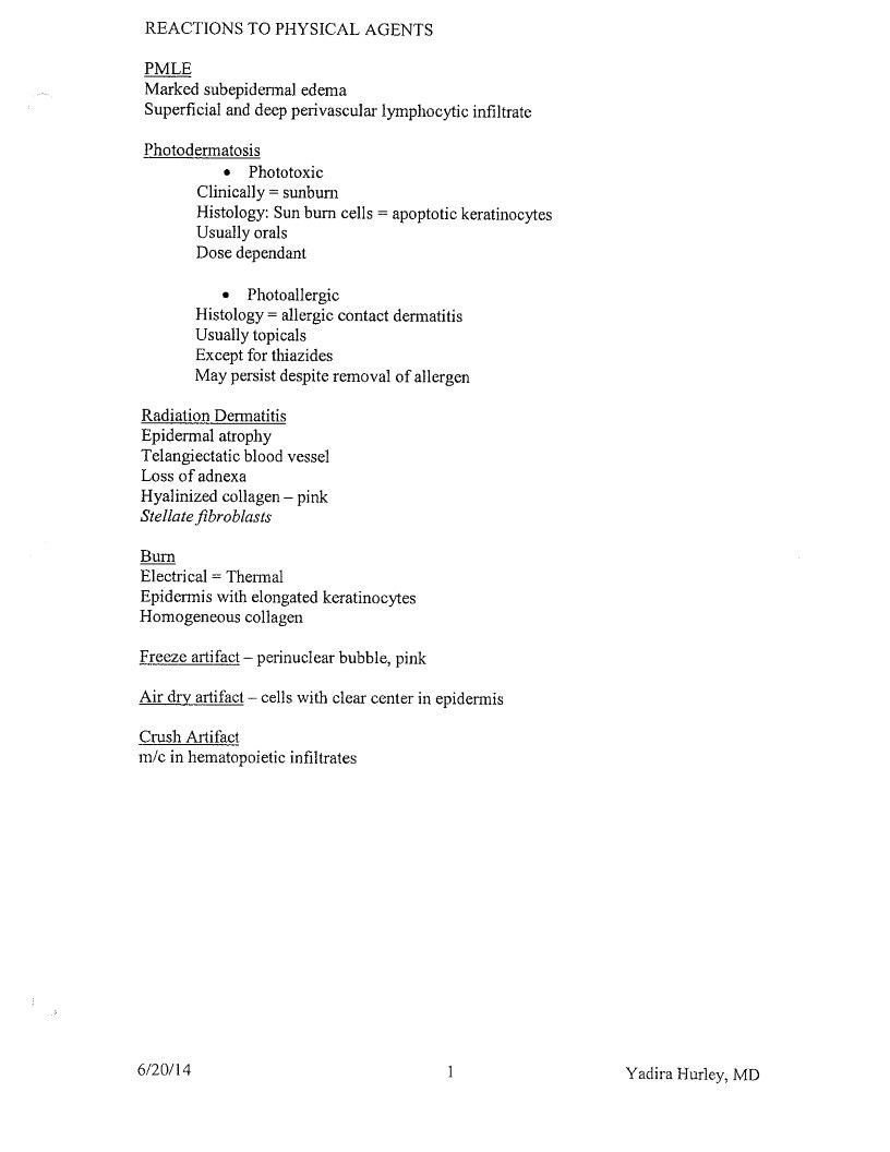
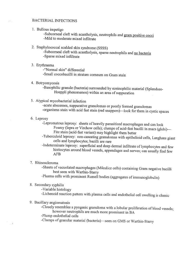
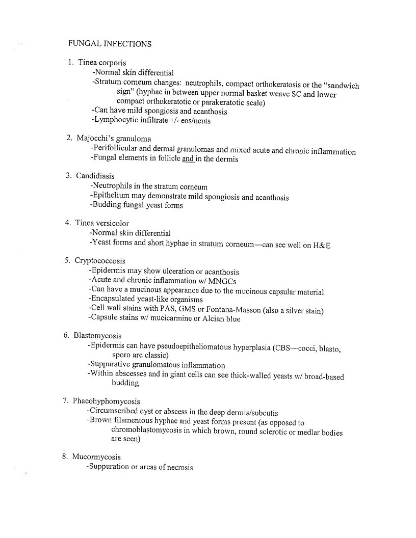
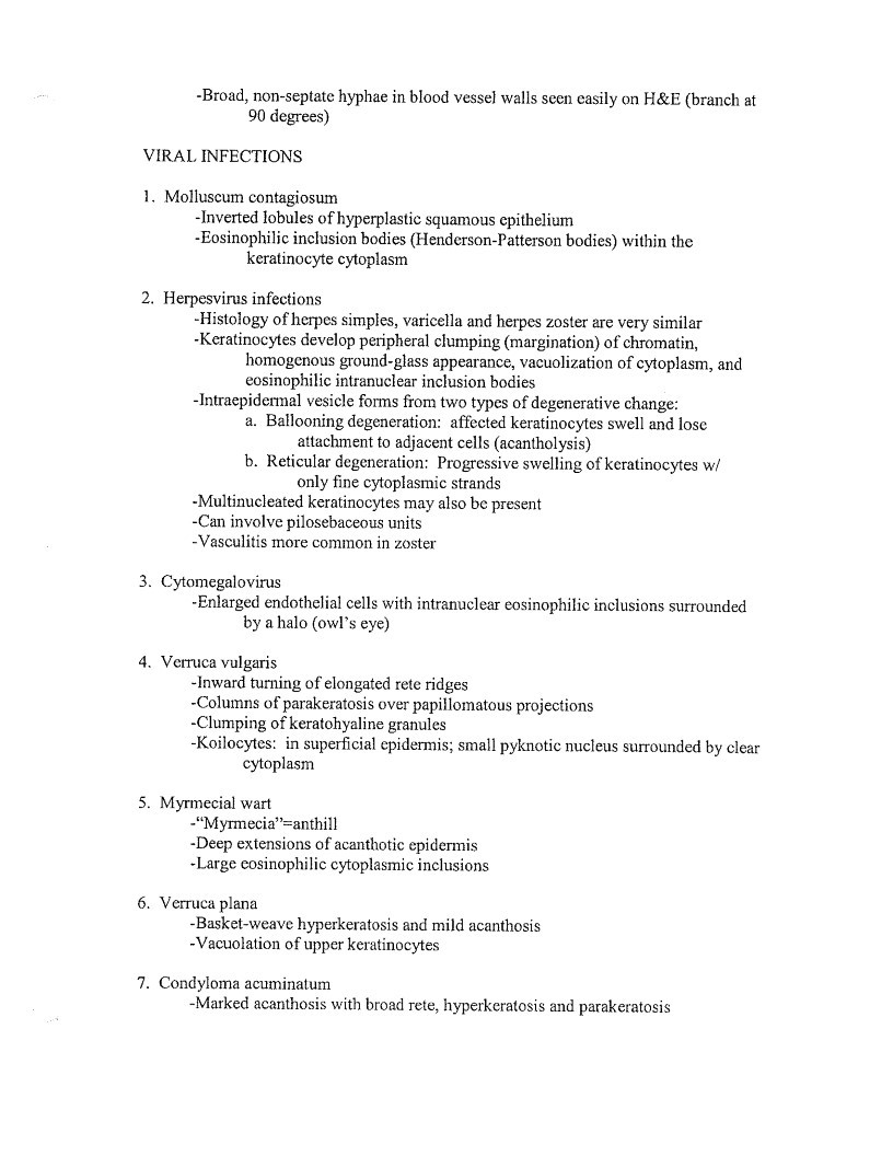
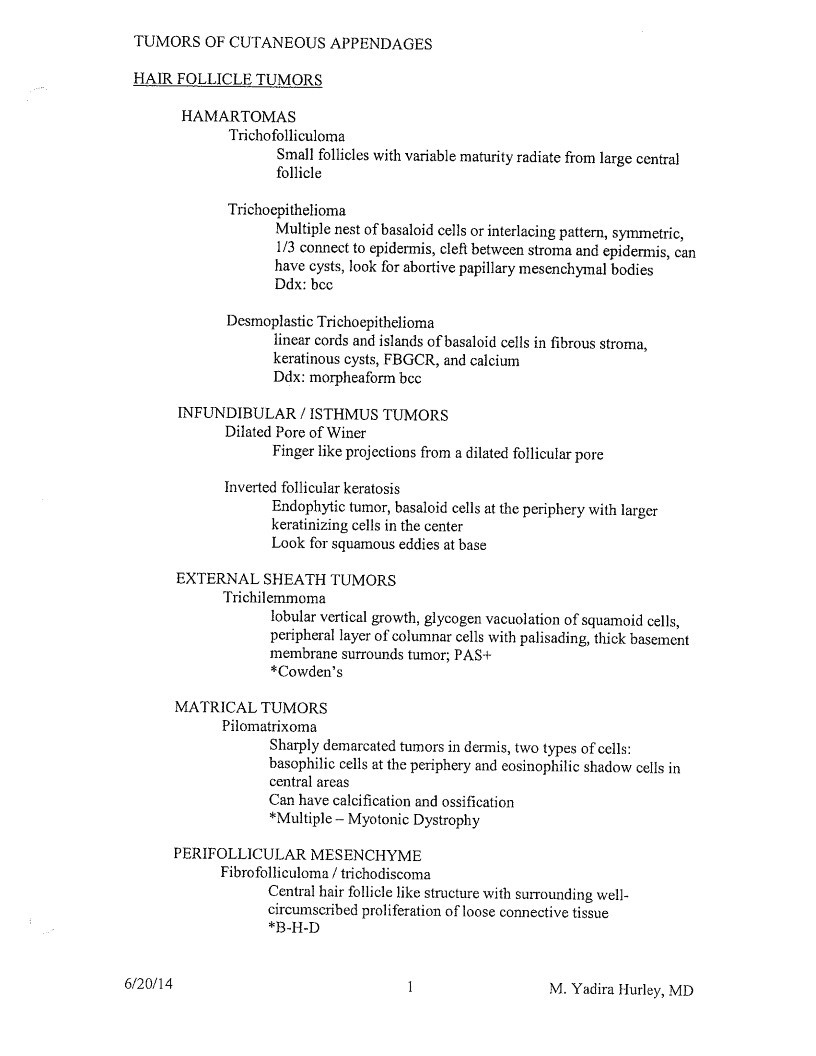
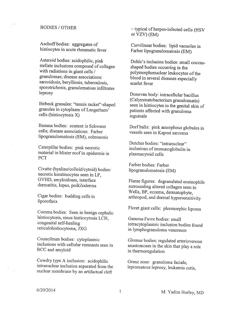
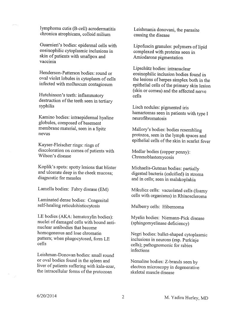
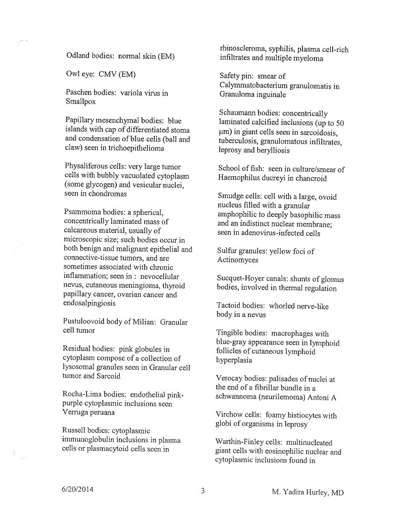
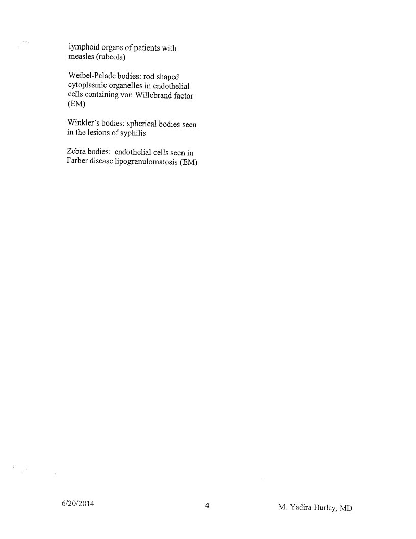
Text from this document (searchable)
Page 1
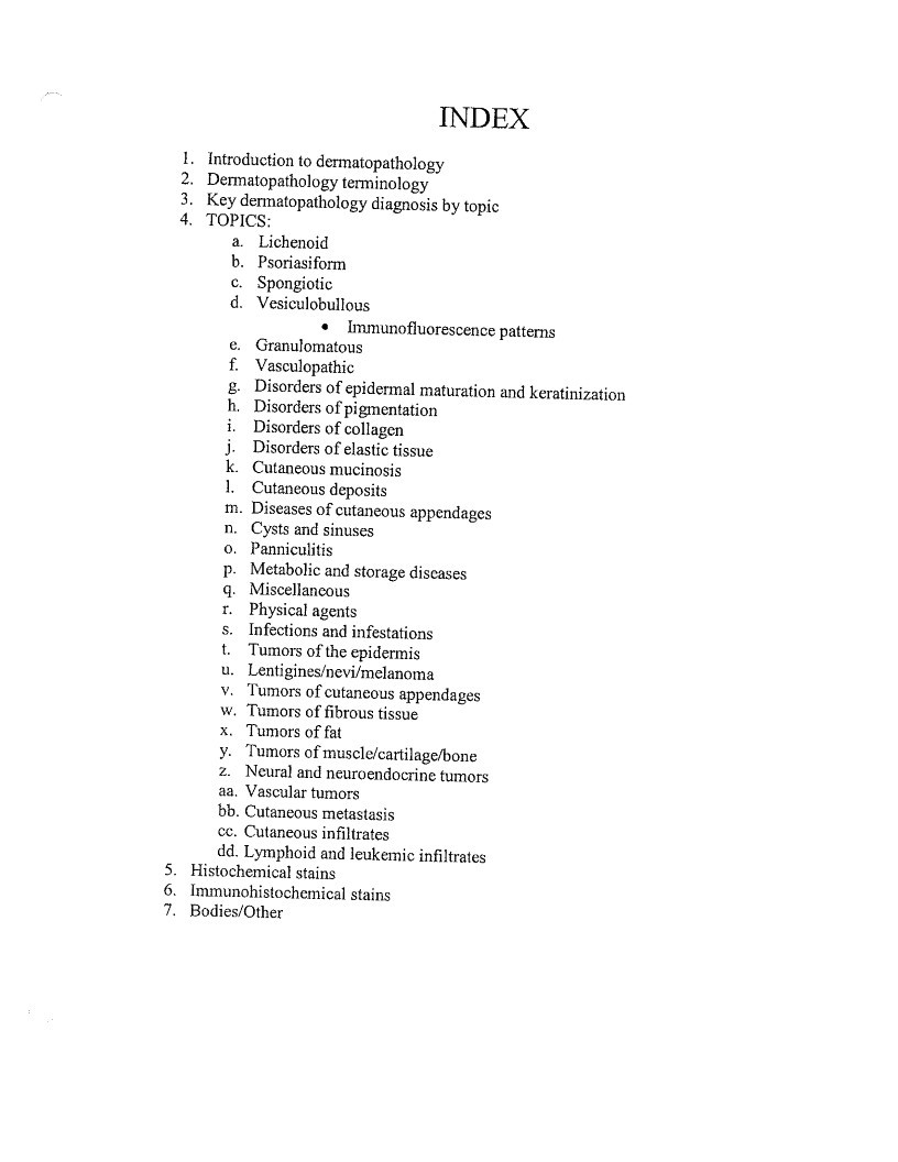
INDEX
- Introduction to dermatopathology
Dermatopathology terminology
Key dermatopathology diagnosis by topic
4.
TOPICS:
a. Lichenoid
b. Psoriasiform
c. Spongiotic
d. Vesiculobullous
• Immunofluorescence patterns
e. Granulomatous
f. Vasculopathic
g. Disorders of epidermal maturation and keratinization
h. Disorders of pigmentation
i. Disorders of collagen
j. Disorders of elastic tissue
k. Cutaneous mucinosis
- Cutaneous deposits
m. Diseases of cutaneous appendages
n. Cysts and sinuses
o. Panniculitis
P. Metabolic and storage discases
q. Miscellaneous
I. Physical agents
s. Infections and infestations
t. Tumors of the epidermis
u. Lentigines/nevi/melanoma
V. Tumors of cutaneous appendages
w. Tumors of fibrous tissue
x. Tumors of fat
y. Tumors of muscle/cartilage/bone
z. Neural and neuroendocrine tumors
aa. Vascular tumors
bb. Cutaneous metastasis
cc. Cutaneous infiltrates
dd. Lymphoid and leukemic infiltrates - Histochemical stains
- Immunohistochemical stains
- Bodies/Other
Page 2
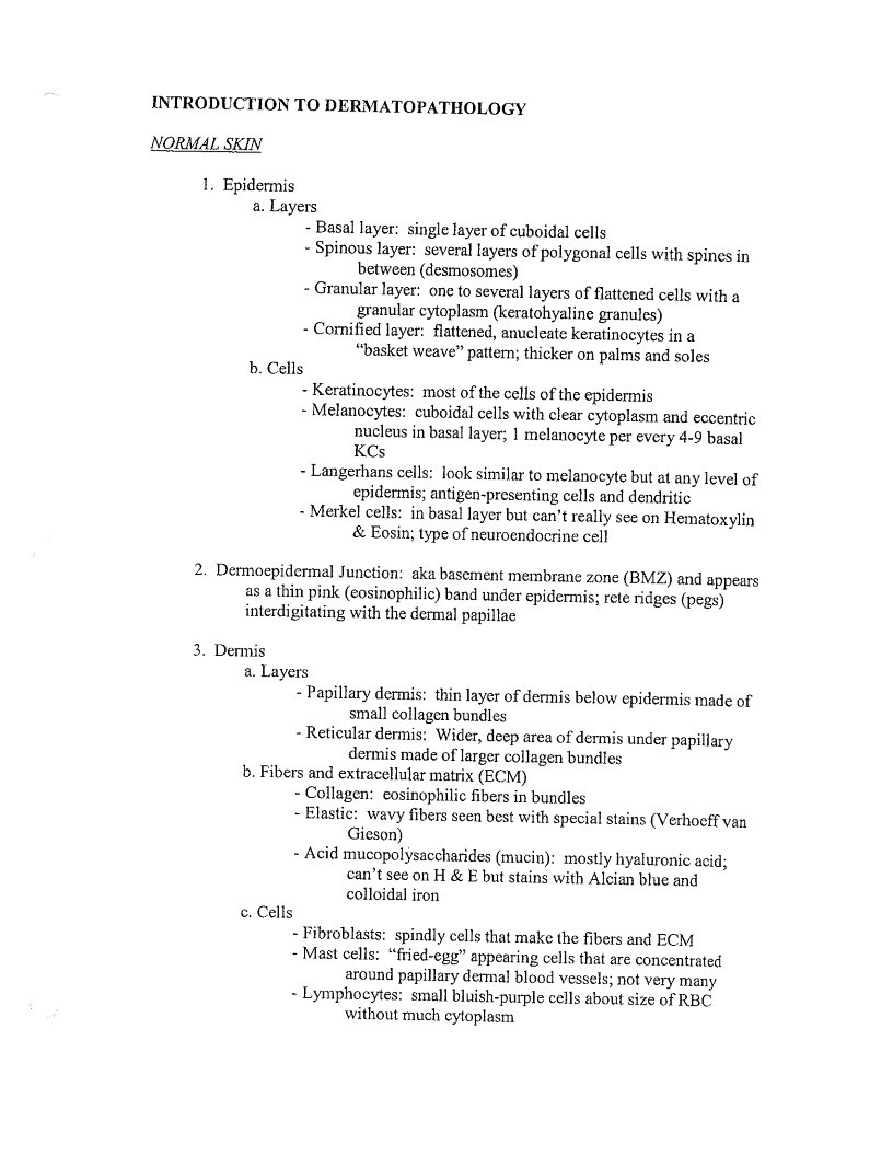
INTRODUCTION TO DERMATOPATHOLOGY
NORMAL SKIN
- Epidermis
a. Layers
- Basal layer: single layer of cuboidal cells
- Spinous layer: several layers of polygonal cells with spines in
between (desmosomes) - Granular layer: one to several layers of flattened cells with a
granular cytoplasm (keratohyaline granules) - Cornified layer: flattened, anucleate keratinocytes in a
”basket weave” pattern; thicker on palms and soles
b. Cells - Keratinocytes: most of the cells of the epidermis
- Melanocytes: cuboidal cells with clear cytoplasm and eccentric
nucleus in basal layer; 1 melanocyte per every 4-9 basal - Langerhans cells: look similar to melanocyte but at any level of
epidermis; antigen-presenting cells and dendritic - Merkel cells: in basal layer but can’t really see on Hematoxylin
& Eosin; type of neuroendocrine cell
- Dermoepidermal Junction: aka basement membrane zone (BMZ) and appears
as a thin pink (eosinophilic) band under epidermis; rete ridges (pegs)
interdigitating with the dermal papillae - Dermis
a. Layers
- Papillary dermis: thin layer of dermis below epidermis made of
small collagen bundles - Reticular dermis: Wider, deep area of dermis under papillary
dermis made of larger collagen bundles
b. Fibers and extracellular matrix (ECM) - Collagen: eosinophilic fibers in bundles
- Elastic: wavy fibers seen best with special stains (Verhoeff van
Gieson) - Acid mucopolýsaccharides (mucin): mostly hyaluronic acid;
can’t see on H & E but stains with Alcian blue and
colloidal iron
c. Cells - Fibroblasts: spindly cells that make the fibers and ECM
- Mast cells: “fried-egg” appearing cells that are concentrated
around papillary dermal blood vessels; not very many - Lymphocytes: small bluish-purple cells about size of RBC
without much cytoplasm
Page 3
- Histiocytes: aka macrophages; mononuclear antigen-presenting
cell with oval, pale-staining nucleus
-Eosinophils: bilobed nucleus and granular cosinophilic
cytoplasm; not usually in normal skin
-Neutrophils: aka polymorphonuclear cells (PMNs);
multilobulated nucleus; not usually in normal skin
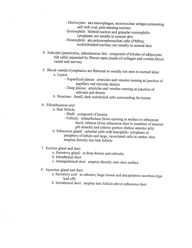
- Subcutis (panniculus, subcutaneous fat): composed of lobules of adipocytes
(fat cells) separated by fibrous septa (made of collagen and contain blood
vessel and nerves) - Blood vessels (lymphatics are flattened so usually not seen in normal skin)
a. Layers
- Superficial plexus: arterioles and venules running at junction of
papillary and reticular dermis - Deep plexus: arterioles and venules running at junction of
subcutis and dermis
b. Structure: Small, dark endothelial cells surrounding the lumen
- Pilosebaceous unit
a. Hair follicle
- Shaft: composed of keratin
- Follicle: infundibulum (from opening at surface to sebaceous
duct), isthmus (from sebaceous duct to insertion of arrector
pili muscle) and inferior portion (below arrector pili)
b. Sebaceous gland: cuboidal cells with basophilic cytoplasm at
periphery of lobule and large, vacuolated cells in center; duct
empties directly into hair follicle
- Eccrine gland and duct
a. Secretory gland: in deep dermis and subcutis
b. Intradermal duct
c. Intrepidermal duct: empties directly onto skin surface - Apocrine gland and duct
a. Secretory coil: in subcutis; large lumen and decapitation secretion (tips
bud off)
b. Intradermal duct: empties into follicle above sebaccous duct
Page 4
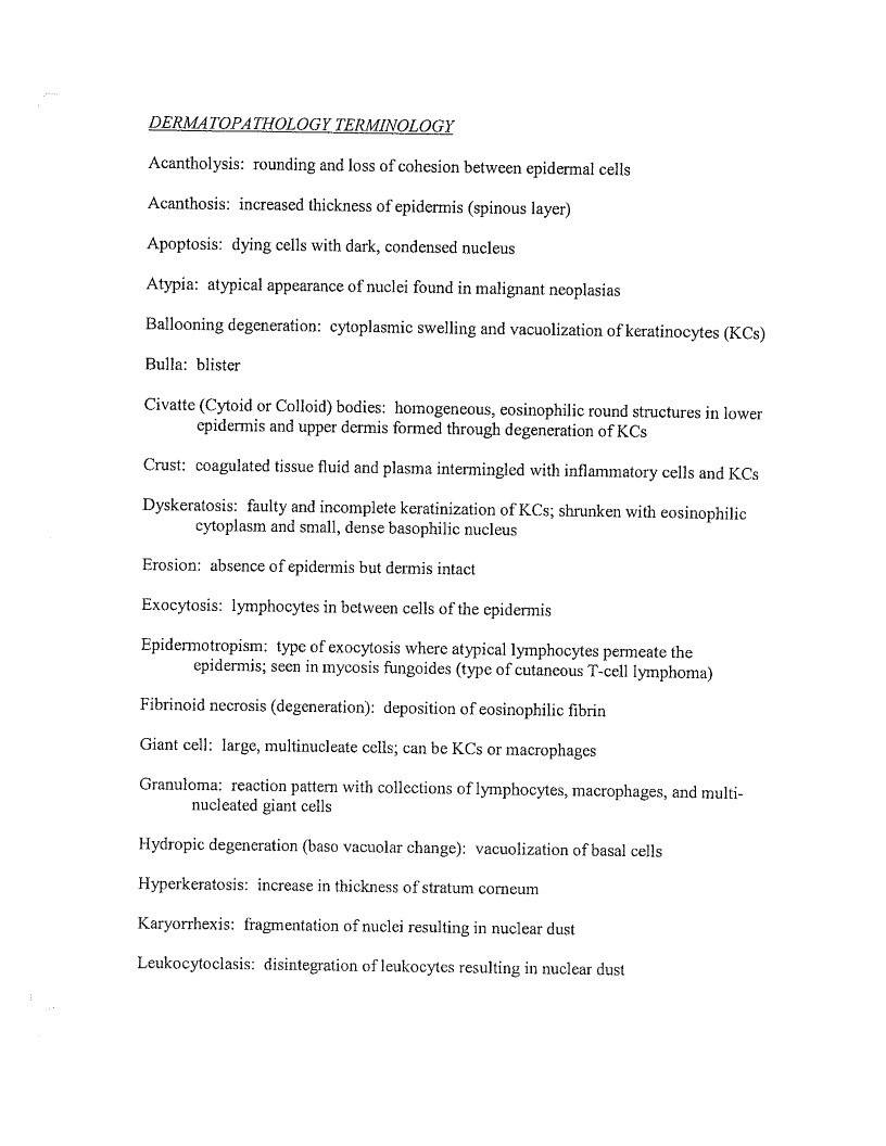
DERMATOPATHOLOGY TERMINOLOGY
Acantholysis: rounding and loss of cohesion between epidermal cells
Acanthosis: increased thickness of epidermis (spinous layer)
Apoptosis: dying cells with dark, condensed nucleus
Atypia: atypical appearance of nuclei found in malignant neoplasias
Ballooning degeneration: cytoplasmic swelling and vacuolization of keratinocytes (KCs)
Bulla: blister
Civatte (Cytoid or Colloid) bodies: homogencous, eosinophilic round structures in lower
cpidermis and upper dermis formed through degeneration of KCs
Crust: coagulated tissue fluid and plasma intermingled with inflammatory cells and KCs
Dyskeratosis: faulty and incomplete keratinization of KCs; shrunken with eosinophilic
cytoplasm and small, dense basophilic nucleus
Erosion: absence of epidermis but dermis intact
Exocytosis: lymphocytes in between cells of the epidermis
Epidermotropism: type of exocytosis where atypical lymphocytes permeate the
epidermis; seen in mycosis fungoides (type of cutaneous T-cell lymphoma)
Fibrinoid necrosis (degeneration): deposition of eosinophilic fibrin
Giant cell: large, multinucleate cells; can be KCs or macrophages
Granuloma: reaction pattern with collections of lymphocytes, macrophages, and multi-
nucleated giant cells
Hydropic degeneration (baso vacuolar change): vacuolization of basal cells
Hyperkeratosis: increase in thickness of stratum corneum
Karyorrhexis: fragmentation of nuclei resulting in nuclear dust
Leukocytoclasis: disintegration of leukocytes resulting in nuclear dust
Page 5
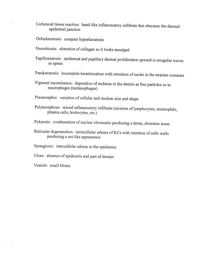
Lichenoid tissue reaction: band-like inflammatory infiltrate that obscures the dermal-
epidermal junction
Orthokeratosis: compact hyperkeratosis
Necrobiosis: alteration of collagen so it looks smudged
Papillomatosis: epidermal and papillary dermal proliferation upward in irregular waves
or spires
Parakeratosis: incomplete keratinization with retention of nuclei in the stratum corneum
Pigment incontinence: deposition of melanin in the dermis as free particles or in
macrophages (melanophages)
Pleomorphic: variation of cellular and nuclear size and shape
Polymorphous: mixed inflammatory infiltrate (mixture of lymphocytes, eosinophils,
plasma cells, histiocytes, etc.)
Pyknosis: condensation of nuclear chromatin producing a dense, shrunken mass
Reticular degeneration: intracellular edema of KCs with retention of cells walls
producing a net-like appearance
Spongiosis: intercellular edema in the cpidermis
Ulcer: absence of epidermis and part of dermis
Vesicle: small blister
Page 6
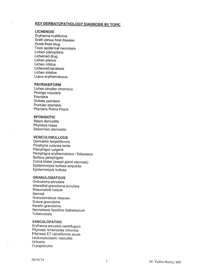
KEY DERMATOPATHOLOGY DIAGNOSIS BY TOPIC
LICHENOID
Erythema multiforme
Graft versus host disease
Acute fixed drug
Toxic epidermal necrolysis
Lichen planopilaris
Lichenoid drug
Lichen planus
Lichen nitidus
Lichenoid keratosis
Lichen striatus
Lupus erythematosus
PSORIASIFORM
Lichen simplex chronicus
Pruirigo nocularis
Psoriasis
Guttate psoriasis
Pustular psoriasis
Pityriasis Rubra Pilaris
SPONGIOTIC
Stasis dermatitis
Pityriasis rosea
Seborrheic dermatitis
VESICULOBULLOUS
Dermatitis herpetiformis
Porphyria cutanea tarda
Pemphigus vulgaris
Pemphigus erythematosus / foliaceous
Bullous pemphigoid
Coma blister (sweat gland necrosis)
Epidermolysis bullosa acquisita
Epidermolysis bullosa
GRANULOMATOUS
Granuloma annulare
Interstitial granuloma annulare
Rheumatoid nodule
Sarcoid
Granulomatous rosacea
Suture granuloma
Keratin granuloma
Necrobiosis lipoidica diabeticorum
Tuberculosis
VASCULOPATHIC
Erythema annulare centrifugum
Pityriasis lichenoides chronica
Pityriasis ET varioliformis acuta
Leukocytoclastic vasculitis
Urticaria
Cryoglobulins
06/20/14
1
M. Yadira Hurley, MID
Page 7
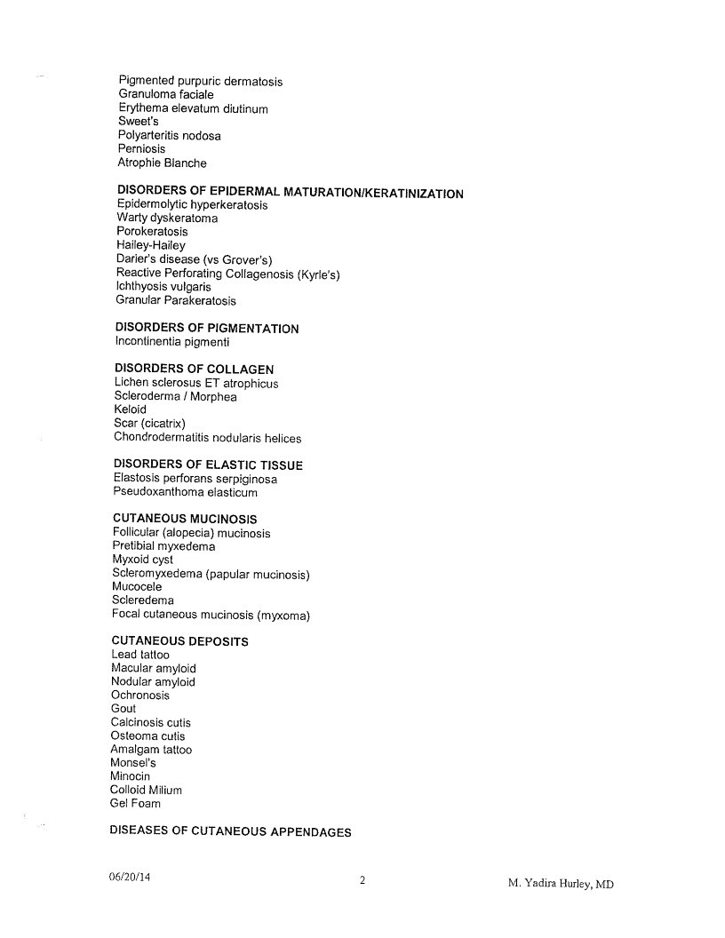
Pigmented purpuric dermatosis
Granuloma faciale
Erythema elevatum diutinum
Sweet’s
Polyarteritis nodosa
Perniosis
Atrophie Blanche
DISORDERS OF EPIDERMAL MATURATION/KERATINIZATION
Epidermolytic hyperkeratosis
Warty dyskeratoma
Porokeratosis
Hailey-Hailey
Darier’s disease (vs Grover’s)
Reactive Perforating Collagenosis (Kyrle’s)
Ichthyosis vulgaris
Granular Parakeratosis
DISORDERS OF PIGMENTATION
Incontinentia pigmenti
DISORDERS OF COLLAGEN
Lichen sclerosus ET atrophicus
Scleroderma / Morphea
Keloid
Scar (cicatrix)
Chondrodermatitis nodularis helices
DISORDERS OF ELASTIC TISSUE
Elastosis perforans serpiginosa
Pseudoxanthoma elasticum
CUTANEOUS MUCINOSIS
Follicular (alopecia) mucinosis
Pretibial myxedema
Myxoid cyst
Scleromyxedema (papular mucinosis)
Mucocele
Scleredema
Focal cutaneous mucinosis (myxoma)
CUTANEOUS DEPOSITS
Lead tattoo
Macular amyloid
Nodular amyloid
Ochronosis
Gout
Calcinosis cutis
Osteoma cutis
Amalgam tattoo
Monsel’s
Minocin
Colloid Milium
Gel Foam
DISEASES OF CUTANEOUS APPENDAGES
06/20/14
2
M. Yadira Hurley, MD
Page 8
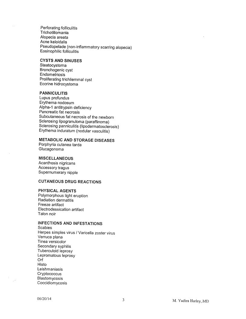
Perforating folliculitis
Trichotillomania
Alopecia areata
Acne keloidalis
Pseudopelade (non-inflammatory scarring alopecia)
Eosinophilic folliculitis
CYSTS AND SINUSES
Steatocystoma
Bronchogenic cyst
Endometriosis
Proliferating trichilemmal cyst
Eccrine hidrocystoma
PANNICULITIS
Lupus profundus
Erythema nodosum
Alpha-1 antitrypsin deficiency
Pancreatic fat necrosis
Subcutaneous fat necrosis of the newborn
Sclerosing lipogranuloma (paraffinoma)
Sclerosing panniculitis (lipodermatosclerosis)
Erythema induratum (nodular vasculitis)
METABOLIC AND STORAGE DISEASES
Porphyria cutanea tarda
Glucagonoma
MISCELLANEOUS
Acanthosis nigricans
Accessory tragus
Supernumerary nipple
CUTANEOUS DRUG REACTIONS
PHYSICAL AGENTS
Polymorphous light eruption
Radiation dermatitis
Freeze artifact
Electrodessication artifact
Talon noir
INFECTIONS AND INFESTATIONS
Scabies
Herpes simples virus / Varicella zoster virus
Verruca plana
Tinea versicolor
Secondary syphilis
Tuberculoid leprosy
Lepromatous leprosy
Orf
Histo
Leishmaniasis
Cryptococcus
Blastomycosis
Coccidiomycosis
06/20/14
3
M. Yadira Hurley, MD
Page 9
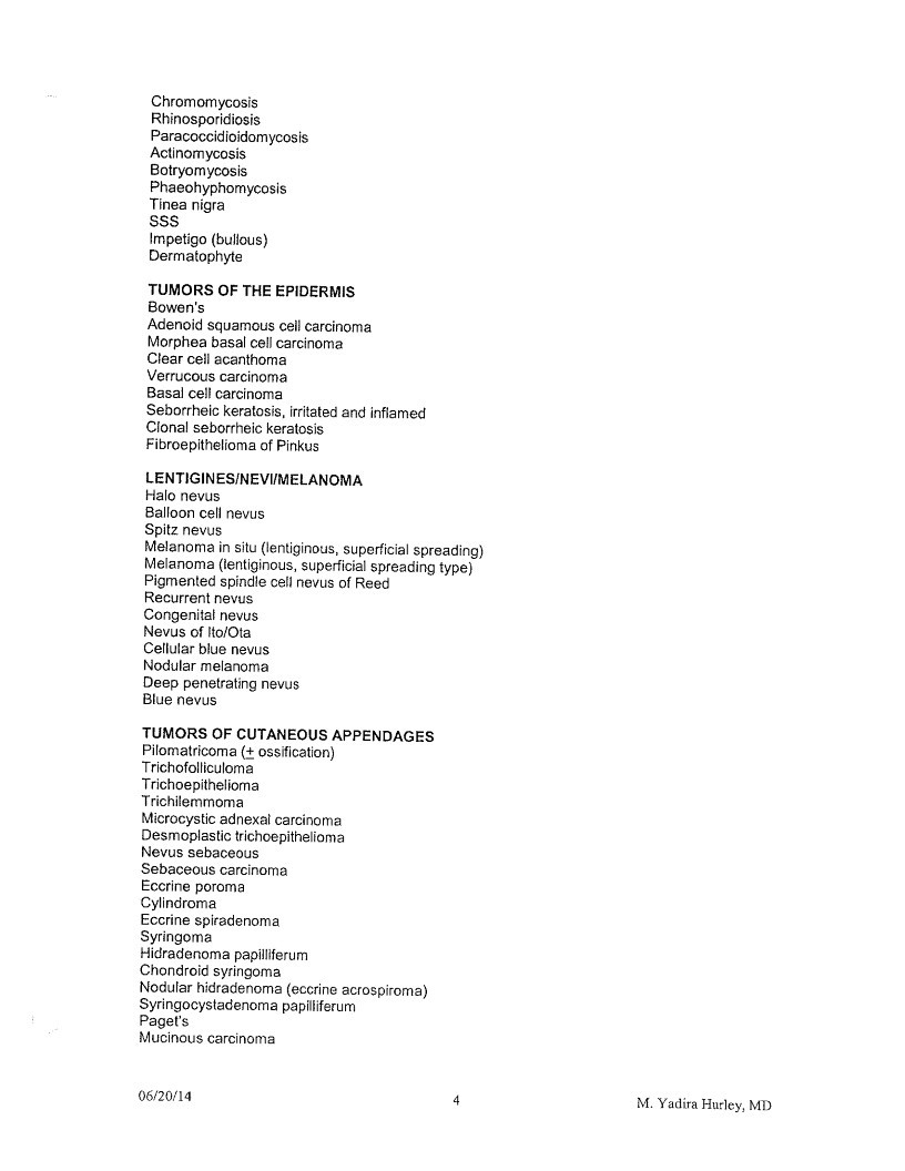
Chromomycosis
Rhinosporidiosis
Paracoccidioidomycosis
Actinomycosis
Botryomycosis
Phaeohyphomycosis
Tinea nigra
SSS
Impetigo (bullous)
Dermatophyte
TUMORS OF THE EPIDERMIS
Bowen’s
Adenoid squamous cell carcinoma
Morphea basal cell carcinoma
Clear cell acanthoma
Verrucous carcinoma
Basal cell carcinoma
Seborrheic keratosis, irritated and inflamed
Clonal seborrheic keratosis
Fibroepithelioma of Pinkus
LENTIGINES/NEVI/MELANOMA
Halo nevus
Balloon cell nevus
Spitz nevus
Melanoma in situ (lentiginous, superficial spreading)
Melanoma (lentiginous, superficial spreading type)
Pigmented spindle cell nevus of Reed
Recurrent nevus
Congenital nevus
Nevus of Ito/Ota
Cellular blue nevus
Nodular melanoma
Deep penetrating nevus
Blue nevus
TUMORS OF CUTANEOUS APPENDAGES
Pilomatricoma (+ ossification)
Trichofolliculoma
Trichoepithelioma
Trichilemmoma
Microcystic adnexal carcinoma
Desmoplastic trichoepithelioma
Nevus sebaceous
Sebaceous carcinoma
Eccrine poroma
Cylindroma
Eccrine spiradenoma
Syringoma
Hidradenoma papilliferum
Chondroid syringoma
Nodular hidradenoma (eccrine acrospiroma)
Syringocystadenoma papilliferum
Paget’s
Mucinous carcinoma
06/20/14
4
M. Yadira Hurley, MD
Page 10
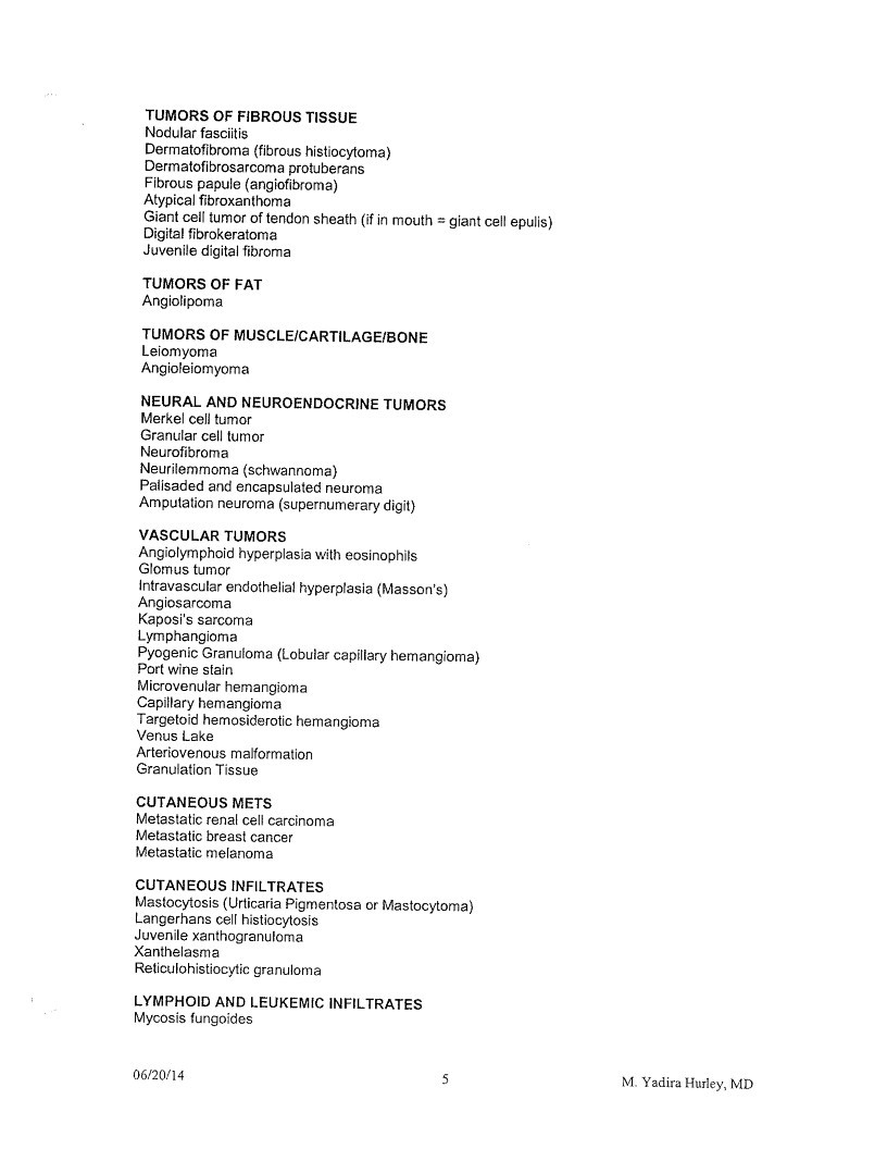
TUMORS OF FIBROUS TISSUE
Nodular fasciitis
Dermatofibroma (fibrous histiocytoma)
Dermatofibrosarcoma protuberans
Fibrous papule (angiofibroma)
Atypical fibroxanthoma
Giant cell tumor of tendon sheath (if in mouth = giant cell epulis)
Digital fibrokeratoma
Juvenile digital fibroma
TUMORS OF FAT
Angiolipoma
TUMORS OF MUSCLE/CARTILAGE/BONE
Leiomyoma
Angioleiomyoma
NEURAL AND NEUROENDOCRINE TUMORS
Merkel cell tumor
Granular cell tumor
Neurofibroma
Neurilemmoma (schwannoma)
Palisaded and encapsulated neuroma
Amputation neuroma (supernumerary digit)
VASCULAR TUMORS
Angiolymphoid hyperplasia with eosinophils
Glomus tumor
Intravascular endothelial hyperplasia (Masson’s)
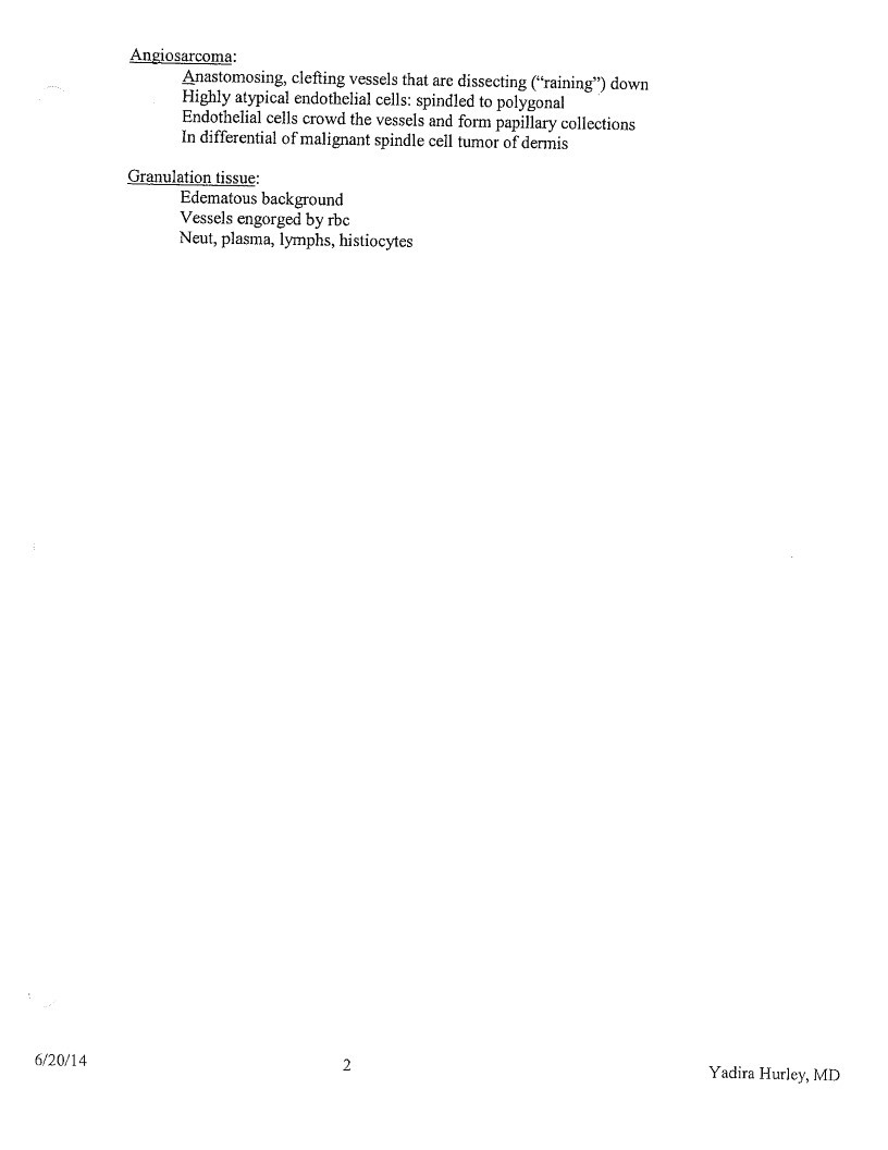
Angiosarcoma
Kaposi’s sarcoma
Lymphangioma
Pyogenic Granufoma (Lobular capillary hemangioma)
Port wine stain
Microvenular hemangioma
Capillary hemangioma
Targetoid hemosiderotic hemangioma
Venus Lake
Arteriovenous malformation
Granulation Tissue
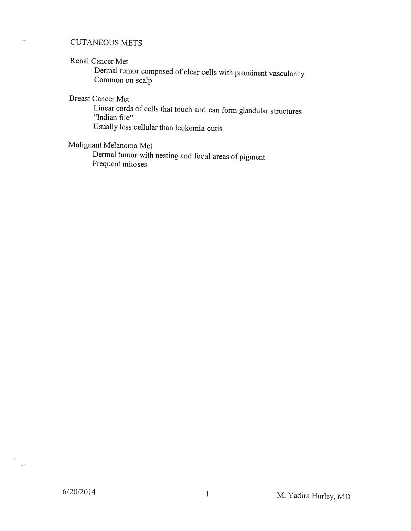
CUTANEOUS METS
Metastatic renal cell carcinoma
Metastatic breast cancer
Metastatic melanoma
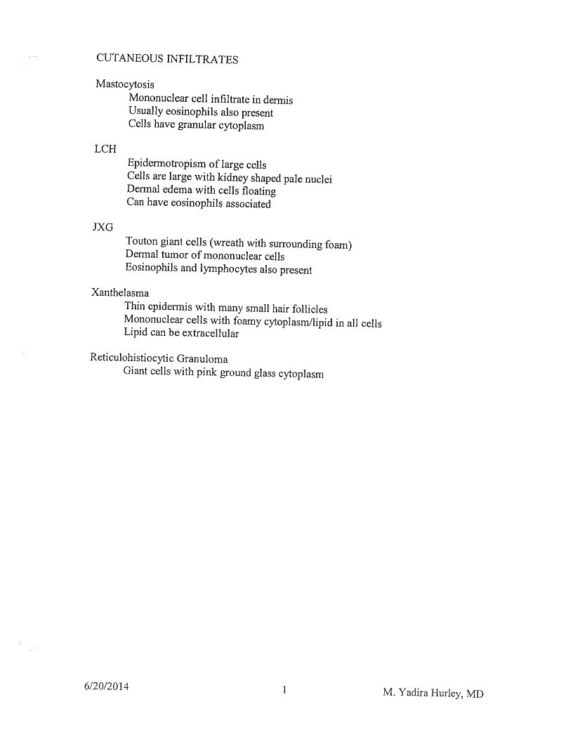
CUTANEOUS INFILTRATES
Mastocytosis (Urticaria Pigmentosa or Mastocytoma)
Langerhans cell histiocytosis
Juvenile xanthogranuloma
Xanthelasma
Reticulohistiocytic granuloma
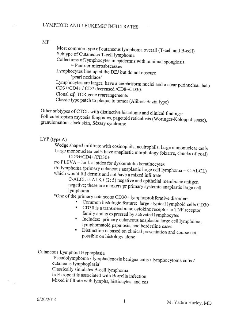
LYMPHOID AND LEUKEMIC INFILTRATES
Mycosis fungoides
06/20/14
5
M. Yadira Hurley, MD
Page 11
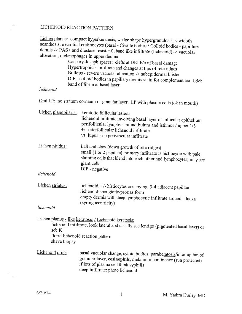
LICHENOID REACTION PATTERN
Lichen planus: compact hyperkeratosis, wedge shape hypergranulosis, sawtooth
acanthosis, necrotic keratinocytes (basal - Civatte bodies / Colloid bodies - papillary
dermis -> PAS+ and diastase resistant), band like infiltrate (lichenoid) -> vacuolar
alteration; melanophages in upper dermis
Caspary-Joseph spaces: clefts at DEJ b/c of basal damage
Hypertrophic - infiltrate and changes at tips of rete ridges
Bullous - severe vacuolar alteration -> subepidermal blister
DIF - colloid bodies in papillary dermis stain for complement and IgM;
band of fibrin at basal layer
lichenoid
Oral LP: no stratum corneum or granular layer. LP with plasma cells (ok in mouth)
Lichen planopilaris:
keratotic follicular lesions
lichenoid infiltrate involving basal layer of follicular epithelium
perifollicular lymphs - infundibulum and isthmus / upper 1/3
+/- interfollicular lichenoid infiltrate
vs. lupus - no perivascular infiltrate
Lichen nitidus:
ball and claw (down growth of rete ridges)
small (1 or 2 papillae), primary infiltrate is histiocytic with pale
staining cells that blend into each other and lymphocytes; may see
giant cells
DIF - negative
lichenoid
Lichen striatus:
lichenoid, +/-histiocytes occupying 3-4 adjacent papillae
lichenoid-spongiotic-psoriasiform
empty dermis with deep lymphocytic infiltrate around adnexa
(syringocentricity)
lichenoid
Lichen piches die kerate, io ident add usualy sce lenigo (pigmented basal layer) o
seb K
florid lichenoid reaction patter
shave biopsy
Lichenoid drug:
basal vacuolar change, cytoid bodies, parakeratosis/interruption of
granular layer, eosinophils, melanin incontinence (sun protected
if lots of plasma cell think syphilis
deep infiltrate: photo lichenoid
6/20/14
1
M. Yadira Hurley, MD
Page 12
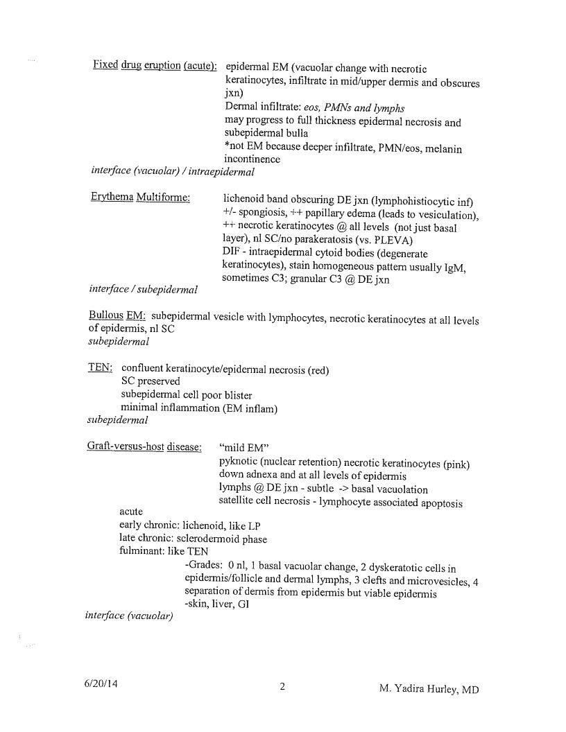
Fixed drug eruption (acute): epidermal EM (vacuolar change with necrotic
keratinocytes, infiltrate in mid/upper dermis and obscures
Dermal infiltrate: eos, PMNs and lymphs
may progress to full thickness epidermal necrosis and
subepidermal bulla
*not EM because decper infiltrate, PM/eos, melanin
incontinence
interface (vacular) / intrapidermal
Erythema Multiforme:
lichenoid band obscuring DE jxn (lymphohistiocytic inf)
+/- spongiosis, ++ papillary edema (leads to vesiculation),
++ necrotic keratinocytes @ all levels (not just basal
layer), ni SC/no parakeratosis (vs. PLEVA
sometimes C3; granular C3 @ DE jxn
interface/subepidermal
Bullous EM: subepidermal vesicle with lymphocytes, necrotic keratinocytes at all levels
of epidermis, n| SC
subepidermal
TEN: confluent keratinocyte/epidernal necrosis (red)
SC preserved
subepidermal cell poor blister
minimal inflammation (EM inflam)
subepidermal
Graft-versus-host disease:
“mild EM”
pyknotic (nuclear retention) necrotic keratinocytes (pink)
down adnexa and at all levels of epidermis
lymphs @ DE ixn - subtle -> basal vacuolation
satellite cell necrosis - lymphocyte associated apoptosis
acute
early chronic: lichenoid, like LP
late chronic: sclerodermoid phase
fulminant: like TEN
-Grades: 0 nl, 1 basal vacuolar change, 2 dyskeratotic cells in
cpidermis/follicle and dermal lymphs, 3 clefts and microvesicles, 4
separation of dermis from epidermis but viable epidermis
-skin, liver, GI
interface (vacuolar)
6/20/14
2
M. Yadira Hurley, MD
Page 13
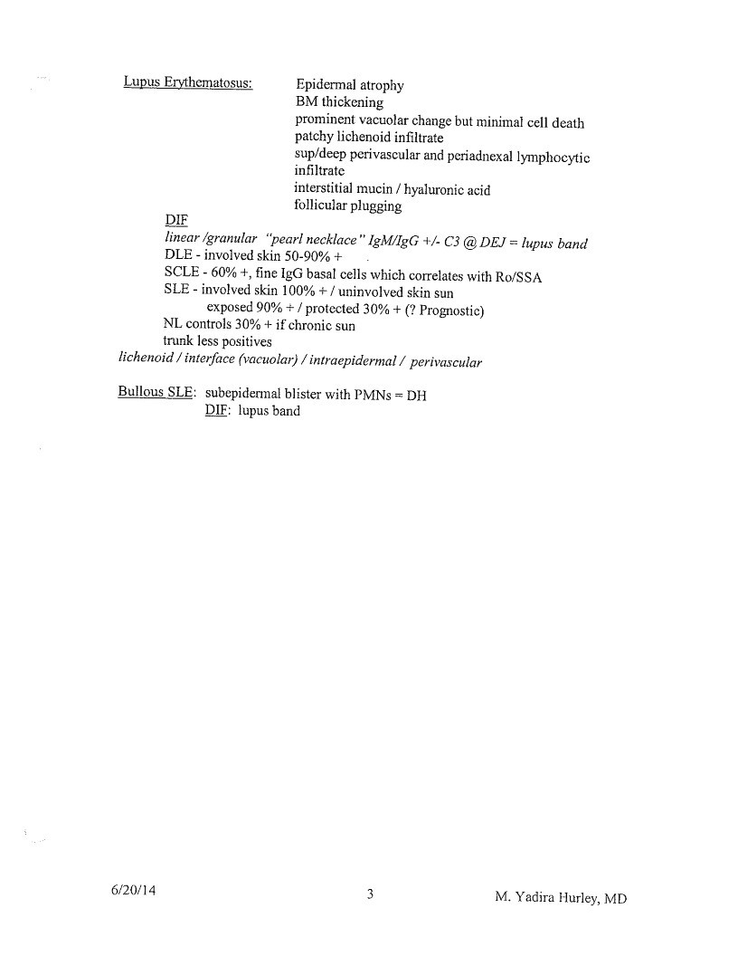
Lupus Erythematosus:
Epidermal atrophy
BM thickening
prominent vacuolar change but minimal cell death
patchy lichenoid infiltrate
sup/deep perivascular and periadnexal lymphocytic
infiltrate
interstitial mucin / hyaluronic acid
follicular plugging
DIF
linear /granular “pearl necklace” IgM/IgG +/ C3 @ DEJ = lupus band
DLE - involved skin 50-90% +
SCLE - 60% +, fine IgG basal cells which correlates with Ro/SSA
SLE - involved skin 100% + / uninvolved skin sun
exposed 90% + / protected 30% + (? Prognostic)
NL controls 30% + if chronic sun
trunk less positives
lichenoid / interface (vacuolar) / intrapidermal / perivascular
Bullous SLE: subepidermal blister with PMNs = DH
DIF: lupus band
6/20/14
3
M. Yadira Hurley, MD
Page 14
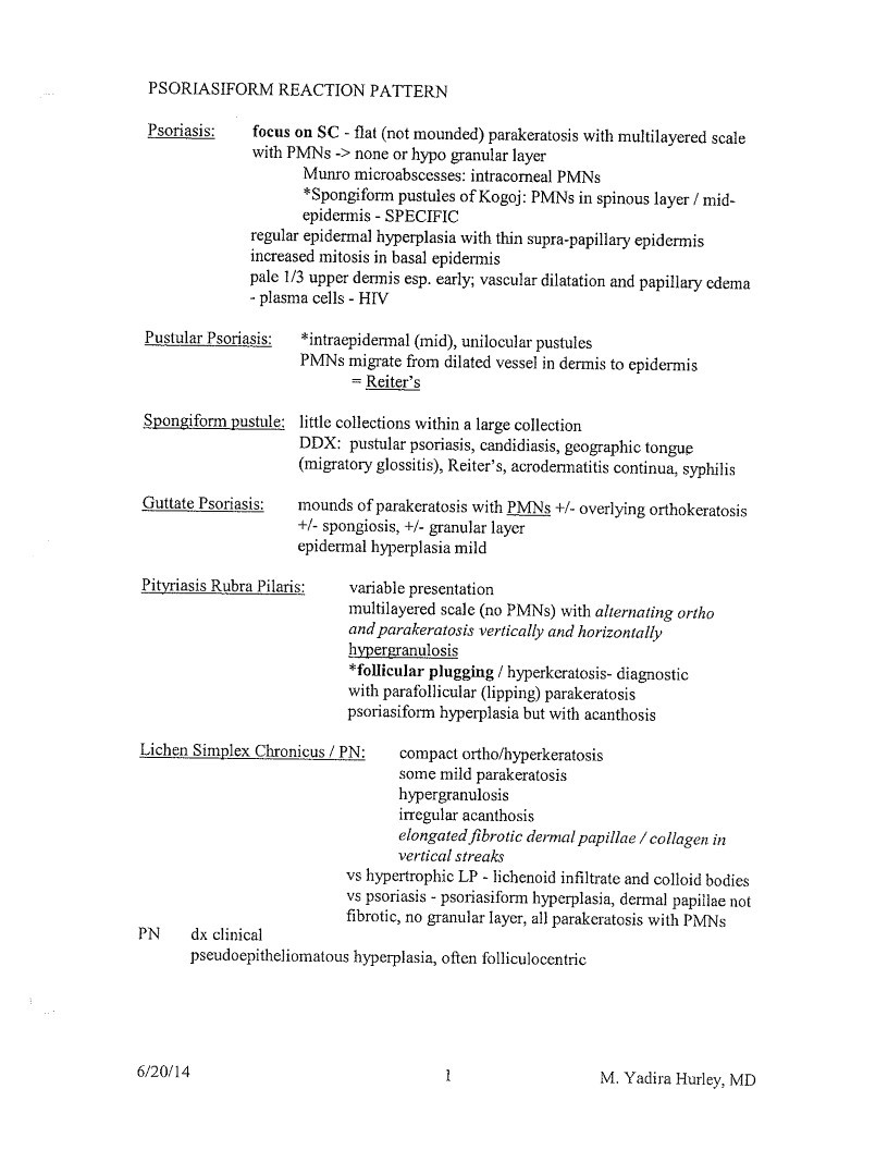
PSORIASIFORM REACTION PATTERN
Psoriasis:
focus on SC - flat (not mounded) parakeratosis with multilayered scale
with PMNs -> none or hypo granular layer
Munro microabscesses: intracorneal PMNs
*Spongiform pustules of Kogoj: PMNs in spinous layer / mid-
epidermis - SPECIFIC
regular epidermal hyperplasia with thin supra-papillary epidermis
increased mitosis in basal epidermis
pale 1/3 upper dermis esp. early; vascular dilatation and papillary dema
- plasma cells - HIV
Pustular Psoriasis:
Spongiform pustule:
*intraepidermal (mid), unilocular pustules
PMNs migrate from dilated vessel in dermis to epidermis
= Reiter’s
little collections within a large collection
DDX: pustular psoriasis, candidiasis, geographic tongue
(migratory glossitis), Reiter’s, acrodermatitis continua, syphilis
Guttate Psoriasis:
mounds of parakeratosis with PMNs +/- overlying orthokeratosis
+/- spongiosis, +/- granular layer
epidermal hyperplasia mild
Pityriasis Rubra Pilaris:
variable presentation
multilayered scale (no PMNs) with alternating ortho
and parakeratosis vertically and horizontally
hypergranulosis
*follicular plugging / hyperkeratosis- diagnostic
with parafollicular (lipping) parakeratosis
psoriasiform hyperplasia but with acanthosis
Lichen Simplex Chronicus / PN:
compact ortho/hyperkeratosis
some mild parakeratosis
hypergranulosis
irregular acanthosis
elongated fibrotic dermal papillae / collagen in
vertical streaks
vs hypertrophic LP - lichenoid infiltrate and colloid bodies
vs psoriasis - psoriasiform hyperplasia, dermal papillae not
fibrotic, no granular layer, all parakeratosis with PMNs
PN
dx clinical
pseudoepitheliomatous hyperplasia, often folliculocentric
6/20/14
1
M. Yadira Hurley, MD
Page 15
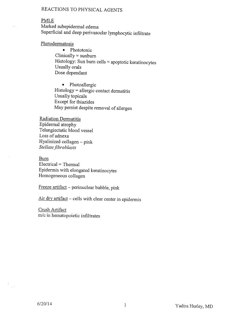
REACTIONS TO PHYSICAL AGENTS
PMLE
Marked subepidermal edema
Superficial and deep perivascular lymphocytic infiltrate
Photodermatosis
• Phototoxic
Clinically = sunburn
Histology: Sun burn cells = apoptotic keratinocytes
Usually orals
Dose dependant
• Photoallergic
Histology = allergic contact dermatitis
Usually topicals
Except for thiazides
May persist despite removal of allergen
Radiation Dermatitis
Epidermal atrophy
Telangiectatic blood vessel
Loss of adnexa
Hyalinized collagen - pink
Stellate fibroblasts
Burn
Electrical = Thermal
Epidermis with elongated keratinocytes
Homogeneous collagen
Freeze artifact - perinuclear bubble, pink
Air dry artifact - cells with clear center in epidermis
Crush Artifact
m/c in hematopoietic infiltrates
6/20/14
1
Yadira Hurley, MD
Page 16
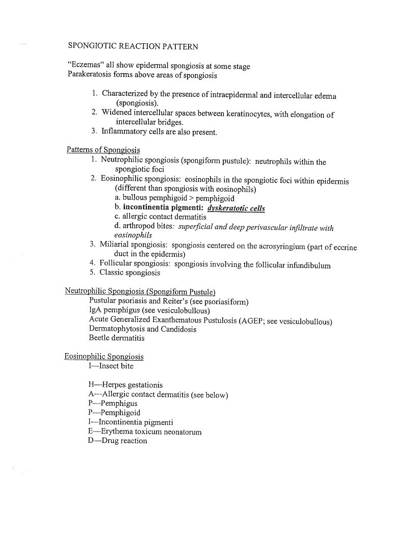
SPONGIOTIC REACTION PATTERN
”Eczemas” all show cpidermal spongiosis at some stage
Parakeratosis forms above areas of spongiosis
- Characterized by the presence of intracpidermal and intercellular edema
(spongiosis). - Widened intercellular spaces between keratinocytes, with elongation of
intercellular bridges. - Inflammatory cells are also present.
Patterns of Spongiosis - Neutrophilic spongiosis (spongiform pustule): neutrophils within the
spongiotic foci - Eosinophilic spongiosis: eosinophils in the spongiotic foci within epidermis
(different than spongiosis with eosinophils)
a. bullous pemphigoid > pemphigoid
b. incontinentia pigmenti: dyskeratotic cells
c. allergic contact dermatitis
d. arthropod bites: superficial and deep perivascular infiltrate with
eosinophils - Miliarial spongiosis: spongiosis centered on the acrosyringium (part of eccrine
duct in the epidermis - Follicular spongiosis: spongiosis involving the follicular infundibulum
- Classic spongiosis
Neutrophilic Spongiosis (Spongiform Pustule)
Pustular psoriasis and Reiter’s (see psoriasiform)
gA pemphigus (see vesiculobullous)
Acute Generalized Exanthematous Pustulosis (AGEP; see vesiculobullous)
Dermatophytosis and Candidosis
Beetle dermatitis
Eosinophilic Spongiosis
1-Insect bite
H-Herpes gestationis
A—Allergic contact dermatitis (see below)
P- Pemphigus
P-Pemphigoid
I-Incontinentia pigmenti
E---Erythema toxicum neonatorum
D—Drug reaction
Page 17
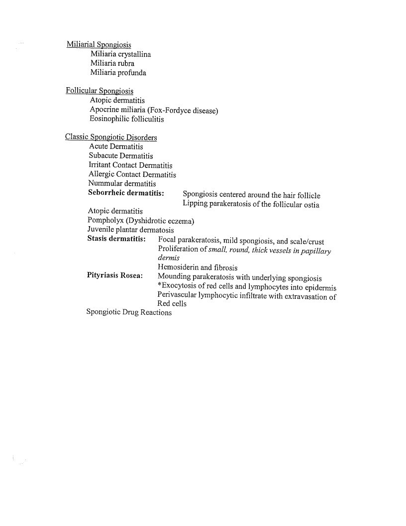
Miliarial Spongiosis
Miliaria crystallina
Miliaria rubra
Miliaria profunda
Follicular Spongiosis
Atopic dermatitis
Apocrine miliaria (Fox-Fordyce disease)
Eosinophilic folliculitis
Classic Spongiotic Disorders
Acute Dermatitis
Subacute Dermatitis
Irritant Contact Dermatitis
Allergic Contact Dermatitis
Nummular dermatitis
Seborrheic dermatitis:
Spongiosis centered around the hair follicle
Lipping parakeratosis of the follicular ostia
Atopic dermatitis
Pompholy (Dyshidrotic eczema)
Juvenile plantar dermatosis
Stasis dermatitis:
Focal parakeratosis, mild spongiosis, and scale/crust
Proliferation of small, round, thick vessels in papillary
dermis
Hemosiderin and fibrosis
Pityriasis Rosea:
Mounding parakeratosis with underlying spongiosis
- Exocytosis of red cells and lymphocytes into epidermis
Perivascular lymphocytic infiltrate with extravasation of
Red cells
Spongiotic Drug Reactions
Page 18
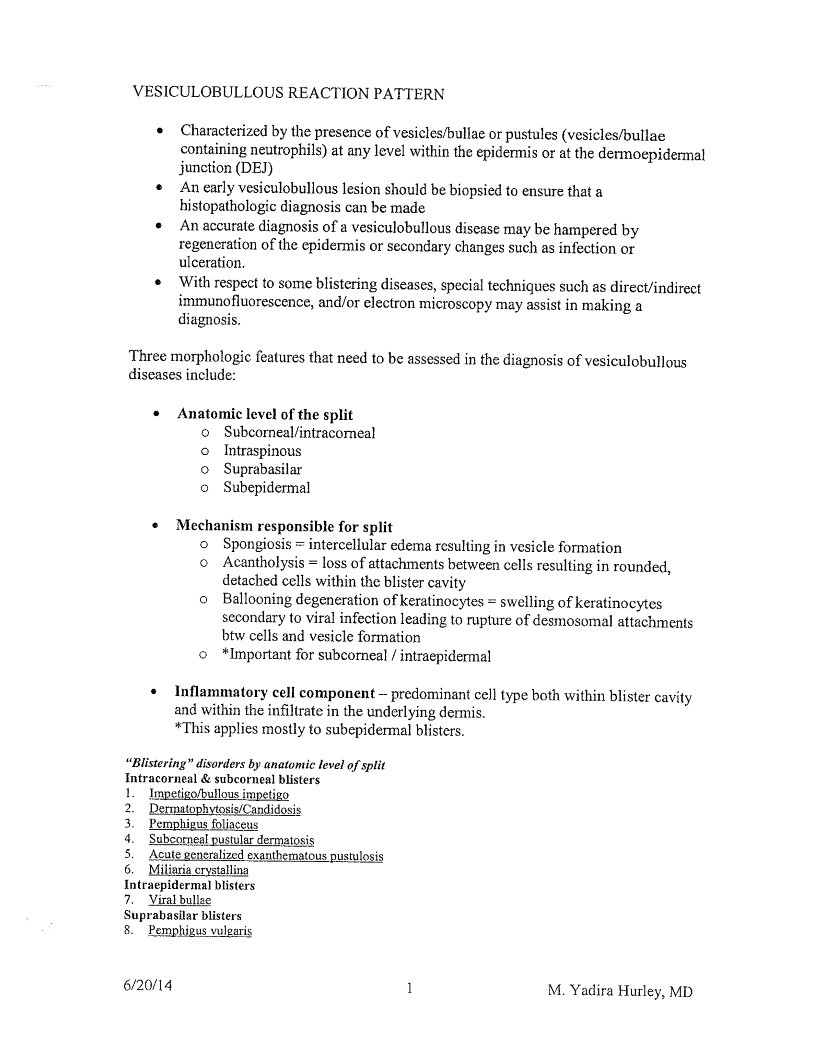
VESICULOBULLOUS REACTION PATTERN
• Characterized by the presence of vesicles/bullae or pustules (vesicles/bullae
containing neutrophils) at any level within the epidermis or at the dermoepidermal
junction (DEJ)
• An early vesiculobullous lesion should be biopsied to ensure that a
histopathologic diagnosis can be made
• An accurate diagnosis of a vesiculobullous disease may be hampered by
regeneration of the epidermis or secondary changes such as infection or
ulceration.
• With respect to some blistering diseases, special techniques such as direct/indirect
immunofluorescence, and/or electron microscopy may assist in making a
diagnosis.
Three morphologic features that need to be assessed in the diagnosis of vesiculobullous
diseases include:
• Anatomic level of the split
Subcorneal/intracomeal
• Intraspinous
Suprabasilar
Subepidermal
• Mechanism responsible for split
Spongiosis = intercellular edema resulting in vesicle formation
• Acantholysis = loss of attachments between cells resulting in rounded,
detached cells within the blister cavity
• Ballooning degeneration of keratinocytes = swelling of keratinocytes
secondary to viral infection leading to rupture of desmosomal attachments
btw cells and vesicle formation
*Important for subcorneal / intraepidermal
• Inflammatory cell component - predominant cell type both within blister cavity
and within the infiltrate in the underlying dermis.
*This applies mostly to subepidermal blisters.
”Blistering” disorders by anatomic level of split
Intracorneal & subcorneal blisters
1.
Impetigo/bullous impetigo
2. Dermatophytosis/Candidosis
3.
Pemphigus foliaceus
5.
Subcorneal pustular dermatosis
Acute generalized exanthematous pustulosis
6. Miliaria crystallina
Intrapidermal blisters
7. Viral bullae
Suprabasilar blisters
Pemphigus vulgaris
6/20/14
M. Yadira Hurley, MD
Page 19
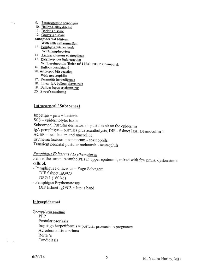
9. Paraneoplastic pemphigus
10. Hailey-Hailey discase
11. Darier’s disease
12. Grover’s disease
Subepidermal blisters:
With little inflammation:
13. Porphyria cutanea tarda
With lymphocytes:
14. Lichen sclerosus et atrophicus
15. Polymorphous light cruption
With eosinophils (Refer to’ I HAPPIED’ mnemonic):
16. Bullous pemphigoid
20. Arthropod bite rcaction
With neutrophils:
17. Dermatitis herpetiformis
18. Linear lgA bullous dermatosis
19. Bullous lupus erythematous
20. Sweet’s syndrome
Intracorneal / Subcorneal
Impetigo - pms + bacteria
SSS - epidermolytic toxin
Subcorneal Pustular dermatosis - pustules sit on the epidermis
IgA pemphigus - pustules plus acantholysis, DIF - fishnet IgA, Desmocollin 1
AGEP - beta lactam and macrolide
Erythema toxicum neonatorum - cosinophils
Transient neonatal pustular melanosis - neutrophils
Pemphigus Foliaceus / Erythematosus
Path is the same: Acantholysis in upper epidermis, mixed with few pmns, dyskeratotic
cells ok
- Pemphigus Foliaceous = Fogo Selvagem
DIF fishnet IgG/C3
DSG 1 (160 kd) - Pemphigus Erythematosus
DIF fishnet IgG/C3 + lupus band
Intraepidermal
Spongiform pustule
PPP
Pustular psoriasis
Impetigo herpetiformis = pustular psoriasis in pregnancy
Acrodermatitis continua
Reiter’s
Candidiasis
6/20/14
2
M. Yadira Hurley, MD
Page 20
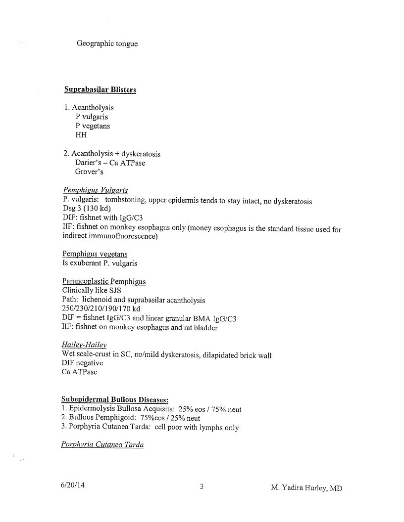
Geographic tongue
Suprabasilar Blisters
- Acantholysis
P vulgaris
P vegetans
HH - Acantholysis + dyskeratosis
Darier’s - Ca ATPasc
Grover’s
Pemphigus Vulgaris
P. vulgaris: tombstoning, upper epidermis tends to stay intact, no dyskeratosis
Dsg 3(130 kd)
IF: fishnet with 1gG/C
IF: fishnet on monkey esophagus only (moncy esophagus is the standard tissue used fo
indirect immunofluorescence)
Pemphigus vegetans
Is exuberant P. vulgaris
Paraneoplastic Pemphigus
Clinically like SJS
Path: lichenoid and suprabasilar acantholysis
250/230/210/190/170 kd
DIF - fishnet IgG/C3 and linear granular BMA IgG/C3
IF: fishnet on monkey esophagus and rat bladder
Hailey-Hailey
Wet scale-crust in SC, no/mild dyskeratosis, dilapidated brick wall
DIF negative
Ca ATPase
Subepidermal Bullous Diseases: - Epidermolysis Bullosa Acquisita: 25% eos / 75% neut
- Bullous Pemphigoid: 75%eos / 25% neut
- Porphyria Cutanea Tarda: cell poor with lymphs only
Porphyria Cutanea Tarda
6/20/14
3
M. Yadira Hurley, MD
Page 21
Subepidermal, lymphs only, thickened BMZ around vessels -> festooning (any
subepidermal blistering disorder can have
Uroporphyrin decarboxylase deficiency
Histologically = pseudo PCT
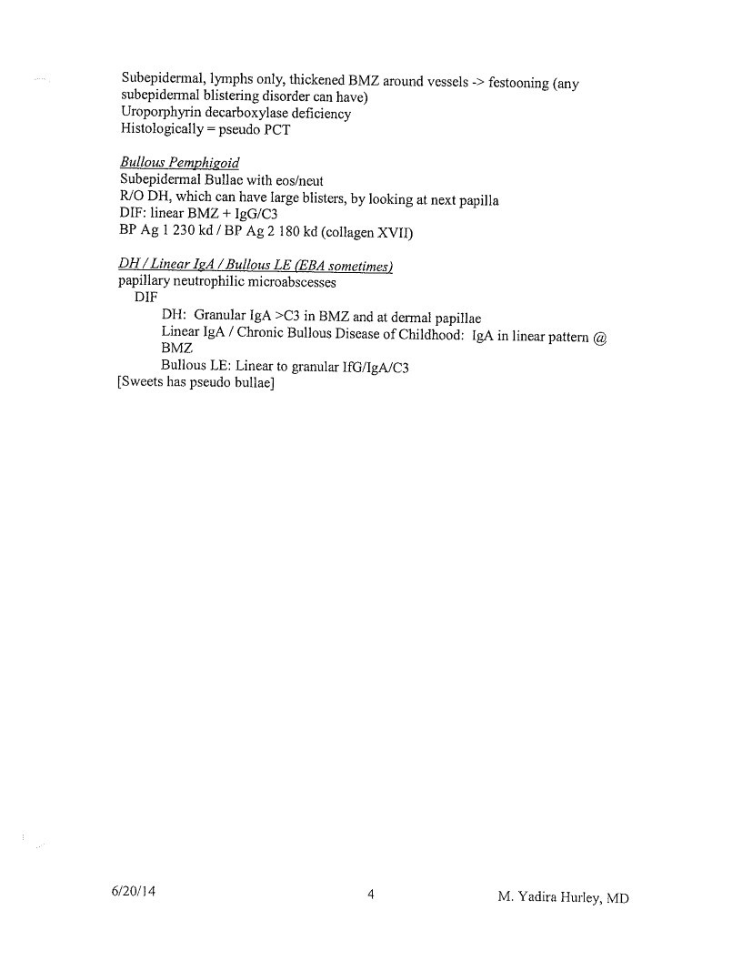
Bullous Pemphigoid
Subepidermal Bullac with eos/neut
R/O DH, which can have large blisters, by looking at next papilla
DIF: linear BMZ + IgG/C3
BP Ag 1 230 kđ / BP Ag 2 180 kd (collagen XVII
DH / Linear IgA / Bullous LE (EBA sometimes)
papillary neutrophilic microabscesses
DIF
DH: Granular IgA >C3 in BMZ and at dermal papillae
Linear IgA / Chronic Bullous Discase of Childhood: IgA in linear pattern @
BMZ.
Bullous LE: Linear to granular IfG/lgA/C3
[Sweets has pseudo bullae]
6/20/14
4
M. Yadira Hurley, MD
Page 22
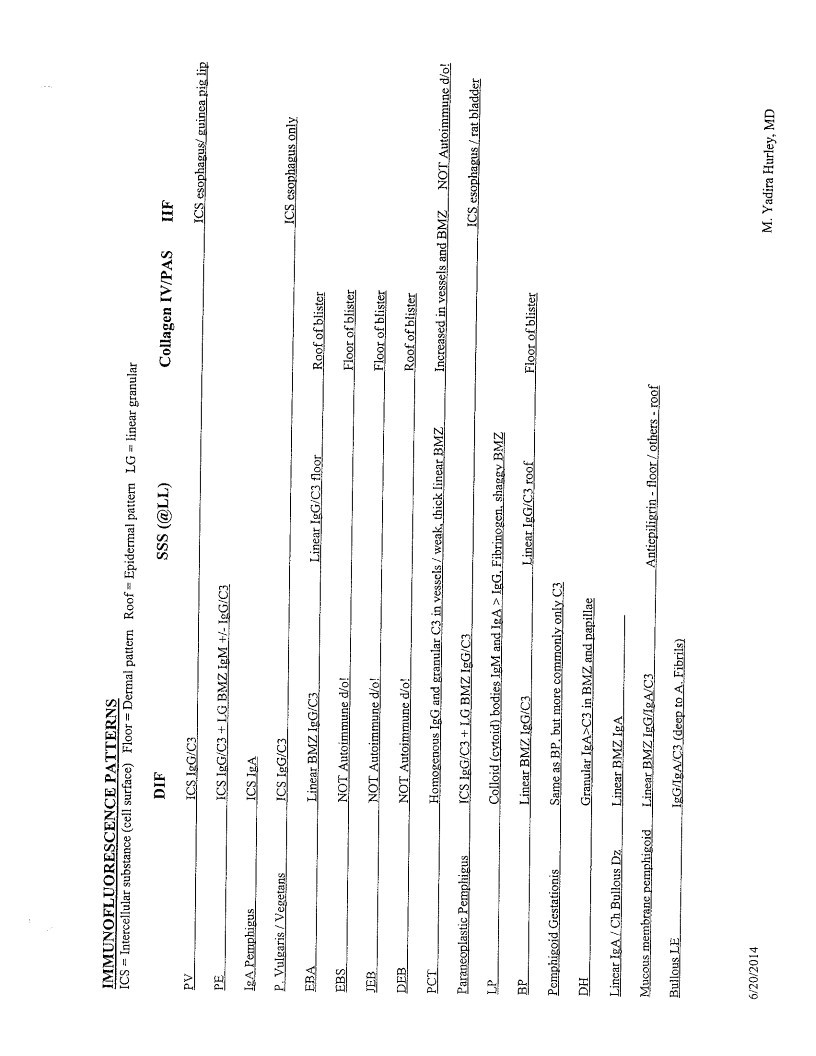
ICs IncreaSES ENCE TO Dermal pater Roof- Epidermal pater 2C - linear granular
SSS (@LL)
IeA. Pemphigus
P. Vulgaris / Vegetans.
EBA.
EBS
JER
DEB
PCT.
Paraneoplastic Pemphigus
Pomphigoid Gestationis
DH
Lincar IgA/Ch Bullous Dz.
Mucous membrane pemphigeid.
Bullous LE
DIF
ICS. IgG/C3
ICS IgG/C3 + LG BMZ IgM +/- IgG/C3
ICS IgA
ICS IgG/C3
Linear BMZ. IG/C3
Linear IgG/C3 floor_
NOT Autoimmune d/o!
NOT Autoimmune dio!
NOT Autoimmune do!
Homogenous IgG and granular C3 in vessels / weak, thick linear BMZ.
ICS IgG/C3 + LG BMZ IgG/C3
Colloid (evoid) bodies IgM and IgA > IgG. Fibrinogen, shaggy BMZ
Linear BMZ. IgG/C3.
Linear IgG/C3 roof_
Same as BP. but more commoniy only C3
Granular IgA>C3 in BMZ and papillae
Linear BMZ IgA.
Linear BMZ IgG/8A/C3
Antiepiligrin - floor / others - roof
IgG/IgA/C3. (deep to A. Fibrils)
Collagen IV/PAS
ICS esophagus/ guinea pig lip
ICS esophagus only
Roof ofblister
Floor of blister
Floor of blister
Roof of blister
Increased in vessels and BMZ NOT Autoimmune do!
ICS esophagus / rat bladder
Floor of blister
6/20/2014
M. Yadira Hurley, MD
Page 23
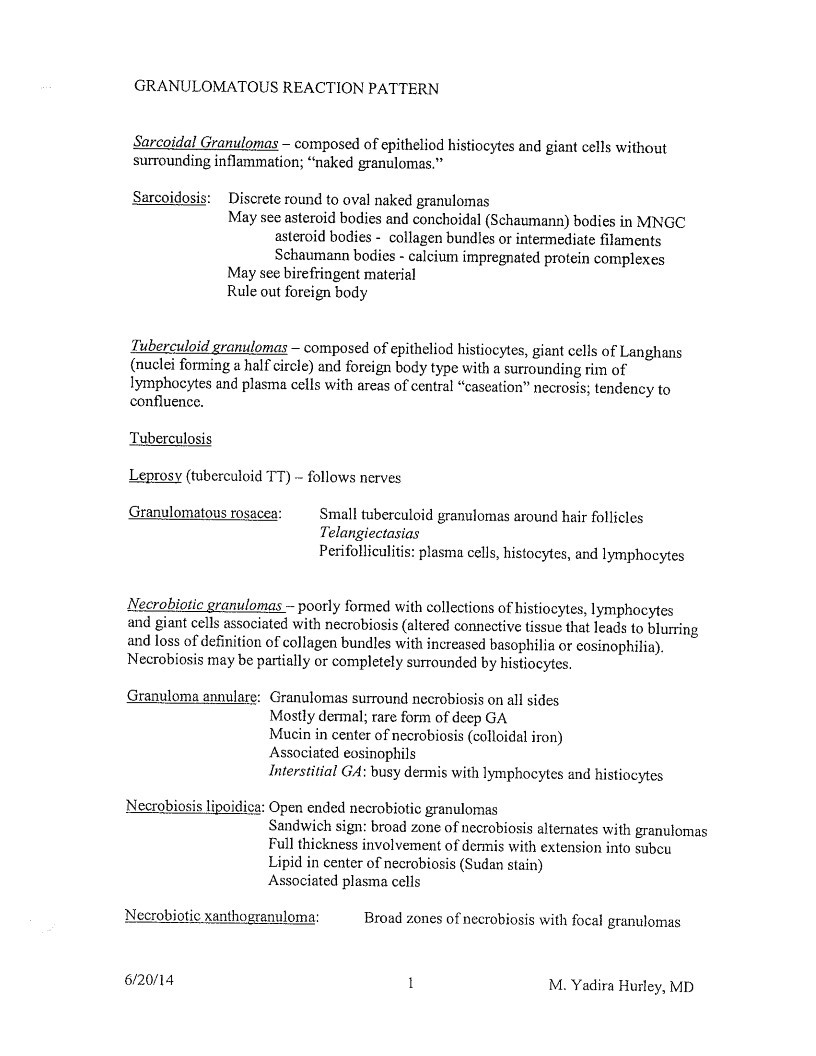
GRANULOMATOUS REACTION PATTERN
Sarcoidal Granulomas - composed of epitheliod histiocytes and giant cells without
surrounding inflammation; “naked granulomas.”
Sarcoidosis:
Discrete round to oval naked granulomas
May see asteroid bodies and conchoidal (Schaumann) bodies in MNGC
asteroid bodies - collagen bundles or intermediate filaments
Schaumann bodies - calcium impregnated protein complexes
May see birefringent material
Rule out foreign body
Tuberculoid granulomas - composed of epitheliod histiocytes, giant cells of Langhans
(nuclei forming a half circle) and foreign body type with a surrounding rim of
lymphocytes and plasma cells with areas of central “caseation” necrosis; tendency to
confluence.
Tuberculosis
Leprosy (tuberculoid TT) - follows nerves
Granulomatous rosacea:
Small tuberculoid granulomas around hair follicles
Telangiectasias
Perifolliculitis: plasma cells, histocytes, and lymphocytes
Necrobiotic granulomas - poorly formed with collections of histiocytes, lymphocytes
and giant cells associated with necrobiosis (altered connective tissue that leads to blurring
and loss of definition of collagen bundles with increased basophilia or cosinophilia).
Necrobiosis may be partially or completely surrounded by histiocytes.
Granuloma annulare: Granulomas surround necrobiosis on all sides
Mostly dermal; rare form of deep GA
Mucin in center of necrobiosis (colloidal iron)
Associated eosinophils
Interstitial GA: busy dermis with lymphocytes and histiocytes
Necrobiosis lipoidica: Open ended necrobiotic granulomas
Sandwich sign: broad zone of necrobiosis alternates with granulomas
Full thickness involvement of dermis with extension into subcu
Lipid in center of necrobiosis (Sudan stain)
Associated plasma cells
Necrobiotic xanthogranuloma:
Broad zones of necrobiosis with focal granulomas
6/20/14
M. Yadira Hurley, MD
Page 24
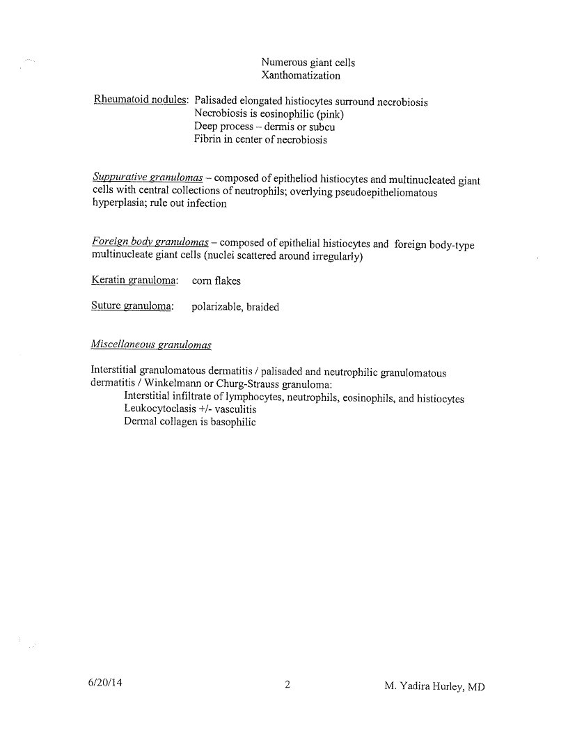
Numerous giant cells
Xanthomatization
Rheumatoid nodules: Palisaded elongated histiocytes surround necrobiosis
Necrobiosis is eosinophilic (pink)
Deep process - dermis or subcu
Fibrin in center of necrobiosis
Suppurative granulomas - composed of epitheliod histiocytes and multinucleated giant
cells with central collections of neutrophils; overlying pseudoepitheliomatous
hyperplasia; rule out infection
Foreign body granulomas - composed of epithelial histiocytes and foreign body-type
multinucleate giant cells (nuclei scattered around irregulariy)
Keratin granuloma:
corn flakes
Suture granuloma:
polarizable, braided
Miscellaneous granulomas
Interstitial granulomatous dermatitis / palisaded and neutrophilic granulomatous
dermatitis / Winkelmann or Churg-Strauss granuloma:
Interstitial infiltrate of lymphocytes, neutrophils, eosinophils, and histiocytes
Leukocytoclasis +/- vasculitis
Dermal collagen is basophilic
6/20/14
2
M. Yadira Hurley, MD
Page 25
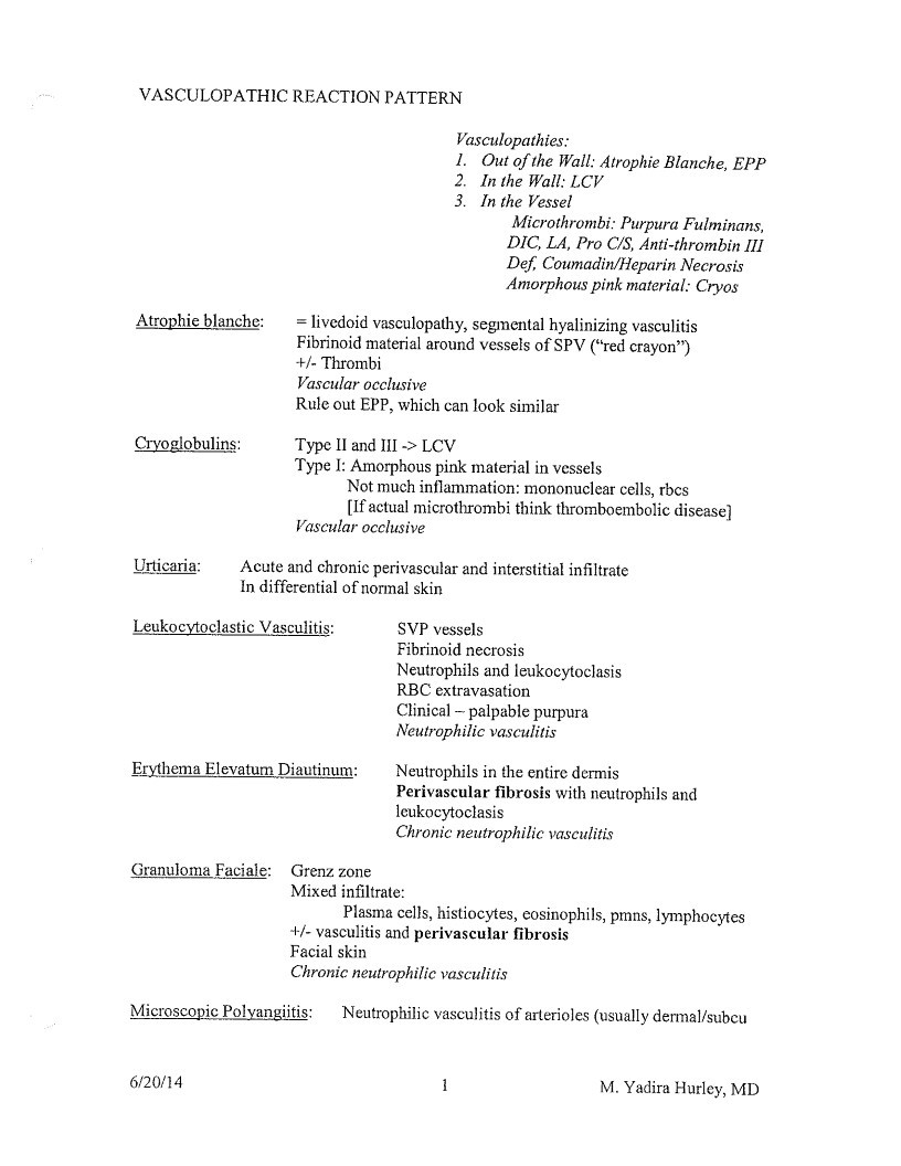
VASCULOPATHIC REACTION PATTERN
Atrophie blanche:
Cryoglobulins:
Vasculopathies:
- Out of the Wall: Atrophie Blanche, EPP
- In the Wall: LCV
- In the Vessel
Microthrombi: Purpura Fulminans,
DIC, LA, Pro C/S, Anti-thrombin III
Def. Coumadin/Heparin Necrosis
Amorphous pink material: Cryos
= livedoid vasculopathy, segmental hyalinizing vasculitis
Fibrinoid material around vessels of SPV (“red crayon”)
+/- Thrombi
Vascular occlusive
Rule out EPP, which can look similar
Type II and III -> LCV
Type I: Amorphous pink material in vessels
Not much inflammation: mononuclear cells, rbcs
[If actual microthrombi think thromboembolic disease?
Vascular occlusive
Urticaria:
Acute and chronic perivascular and interstitial infiltrate
In differential of normal skin
Leukocytoclastic Vasculitis:
SVP vessels
Fibrinoid necrosis
Neutrophils and leukocytoclasis
RBC extravasation
Clinical - palpable purpura
Neutrophilic vasculitis
Erythema Elevatum Diautinum:
Neutrophils in the entire dermis
Perivascular fibrosis with neutrophils and
leukocytoclasis
Chronic neutrophilic vasculitis
Granuloma Faciale:
Grenz zone
Mixed infiltrate:
Plasma cells, histiocytes, eosinophils, pmns, lymphocytes
+/- vasculitis and perivascular fibrosis
Facial skin
Chronic neutrophilic vasculitis
Microscopic Polyangiitis:
Neutrophilic vasculitis of arterioles (usually dermal/subcu
6/20/14
1
M. Yadira Hurley, MD
Page 26
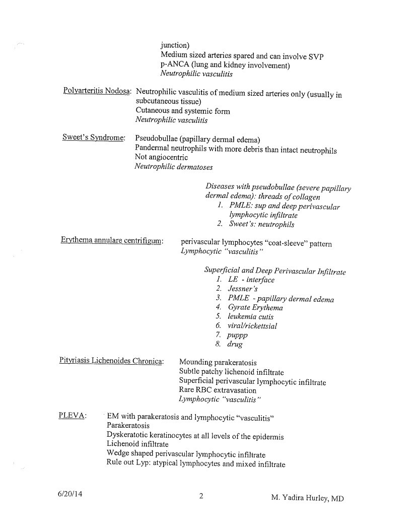
junction)
Medium sized arteries spared and can involve SVP
p-ANCA (lung and kidney involvement)
Neutrophilic vasculitis
Polyarteritis Nodosa: Neutrophilic vasculitis of medium sized arteries only (usually in
subcutaneous tissue)
Cutaneous and systemic form
Neutrophilic vasculitis
Sweet’s Syndrome:
Pseudobullae (papillary dermal edema)
Panderal neutrophils with more debris than intact neutrophils
Not angiocentric
Neutrophilic dermatoses
Diseases with pseudobullae (severe papillary
dermal edema): threads of collagen
- PMLE: sup and deep perivascular
lymphocytic infiltrate - Sweet’s: neutrophils
Erythema annulare centrifigum:
perivascular lymphocytes “coat-sleeve” pattern
Lymphocytic “vasculitis”
Superficial and Deep Perivascular Infiltrate - LE - interface
- Jessner’s
PMLE - papillary dermal edema - Gyrate Erythema
- leukemia cutis
- viral/rickettsial
• puppp
8. drug
Pityriasis Lichenoides Chronica:
Mounding parakeratosis
Subtle patchy lichenoid infiltrate
Superficial perivascular lymphocytic infiltrate
Rare RBC extravasation
Lymphocytic “vasculitis”
PLEVA:
EM with parakeratosis and lymphocytic “vasculitis”
Parakeratosis
Dyskeratotic keratinocytes at all levels of the epidermis
Lichenoid infiltrate
Wedge shaped perivascular lymphocytic infiltrate
Rule out Lyp: atypical lymphocytes and mixed infiltrate
6/20/14
2
M. Yadira Hurley, MD
Page 27
Lymphocytic “vasculitis”
Pigmented Purpuric Dermatoses:
Capilliritis (lymphocytes and rbc extravasation)
Hemosiderin
Perniosis:
Acral skin
Superficial and deep perivascular lymphocytic infiltrate
+/- interface change
Lymphocytic “vasculitis”
6/20/14
3
M. Yadira Hurley, MD
Page 28
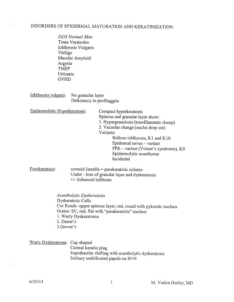
DISORDERS OF EPIDERMAL MATURATION AND KERATINIZATION
DDX Normal Skin:
Tinea Versicolor
Ichthyosis Vulgaris
Vitiligo
Macular Amyloid
Argyria
Urticaria
GVHD
Ichthyosis vulgaris:
No granular layer
Deficiency in profilaggrin
Epidermolytic Hyperkeratosis:
Compact hyperkeratosis
Spinous and granular layer show:
- Hypergranulosis (tonofilaments clump)
- Vacuolar change (nuclei drop out)
Variants:
Bullous ichthyosis, K1 and K10
Epidermal nevus - variant
PPK - variant (Vorner’s syndrome), K9
Epidermolytic acanthoma
Incidental
Porokeratosis:
cornoid lamella = parakeratotic column
Under - loss of granular layer and dyskeratosis
+/- lichenoid infiltrate
Acantholytic Dyskeratosis
Dyskeratotic Cells
or Ronds: upper spinous layer; red, round with pyknotic nucleu
irains: SC; red, flat with “parakeratotic” nucleu - Warty Dyskeratoma
- Darier’s
- Grover’s
Warty Dyskeratoma: Cup shaped
Central keratin plug
Suprabasilar clefting with acantholytic dyskeratosis
Solitary umbilicated papule on H+N
6/20/14
1
M. Yadira Huricy, MD
Page 29
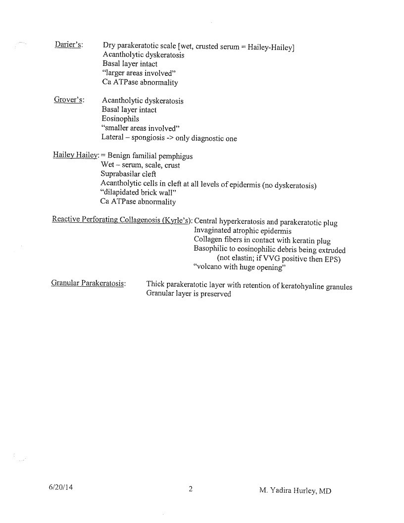
Darier’s:
Dry parakeratotic scale [wet, crusted serum = Hailey Hailey]
Acantholytic dyskeratosis
Basal layer intact
”larger areas involved”
Ca ATPase abnormality
Grover’s:
A cantholytic dyskeratosis
Basal layer intact
Eosinophils
”smaller areas involved”
Lateral - spongiosis -> only diagnostic one
Hailey Hailey: = Benign familial pemphigus
Wet - serum, scale, crust
Suprabasilar cleft
Acantholytic cells in cleft at all levels of epidermis (no dyskeratosis)
“dilapidated brick wall”
Ca ATPase abnormality
Reactive Perforating Collagenosis (Kyrle’s): Central hyperkeratosis and parakeratotic plug
Invaginated atrophic epidermis
Collagen fibers in contact with keratin plug
Basophilic to eosinophilic debris being extruded
(not elastin; if VVG positive then EPS)
“volcano with huge opening”
Granular Parakeratosis:
Thick parakeratotic layer with retention of keratohyaline granules
Granular layer is preserved
6/20/14
2
M. Yadira Hurley, MD
Page 30
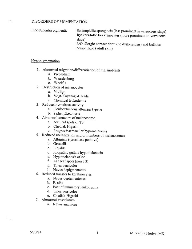
DISORDERS OF PIGMENTATION
Incontinentia pigmenti:
Eosinophilic spongiosis (less prominent in verrucous stage)
Dyskeratotic keratinocytes (more prominent in verrucous
stage)
R/O allergic contact derin (no dyskeratosis) and bullous
pemphigoid (adult skin)
Hypopigmentation
- Abnormal migration/differentiation of melanoblasts
a. Piebaldism
b. Waardenburg
C.
Woolf s - Destruction of melanocytes
a.
Vitiligo
b.
Vogt-Koyanagi-Harada
c. Chemical leukoderma - Reduced tyrosinase activity
a. Oculocutaneous albinism type A
b. ? phenylketonuria - Abnormal structure of melanosome
a. Ash leaf spots of TS
b. Chediak-Higashi
c. Progressive macular hypomelanosis - Reduced melanization and/or numbers of melanosomes
a. Albinism (tyrosinase positive)
b. Griscelli
c. Elejalde
d. Idiopathic guttate hypomelanosis
e. Hypomelanosis of Ito
f. Ash leaf spots (non TS)
g. Tinea versicolor
h. Nevus depigmentosus - Reduced transfer to keratinocytes
a. Nevus depigmentosus
b. P. alba
c. Postinflammatory leukoderma
d. Tinea versicolor
e. Chediak-Higashi - Abnormal vasculature
a. Nevus anemicus
6/20/14
M. Yadira Hurley, MD
Page 31
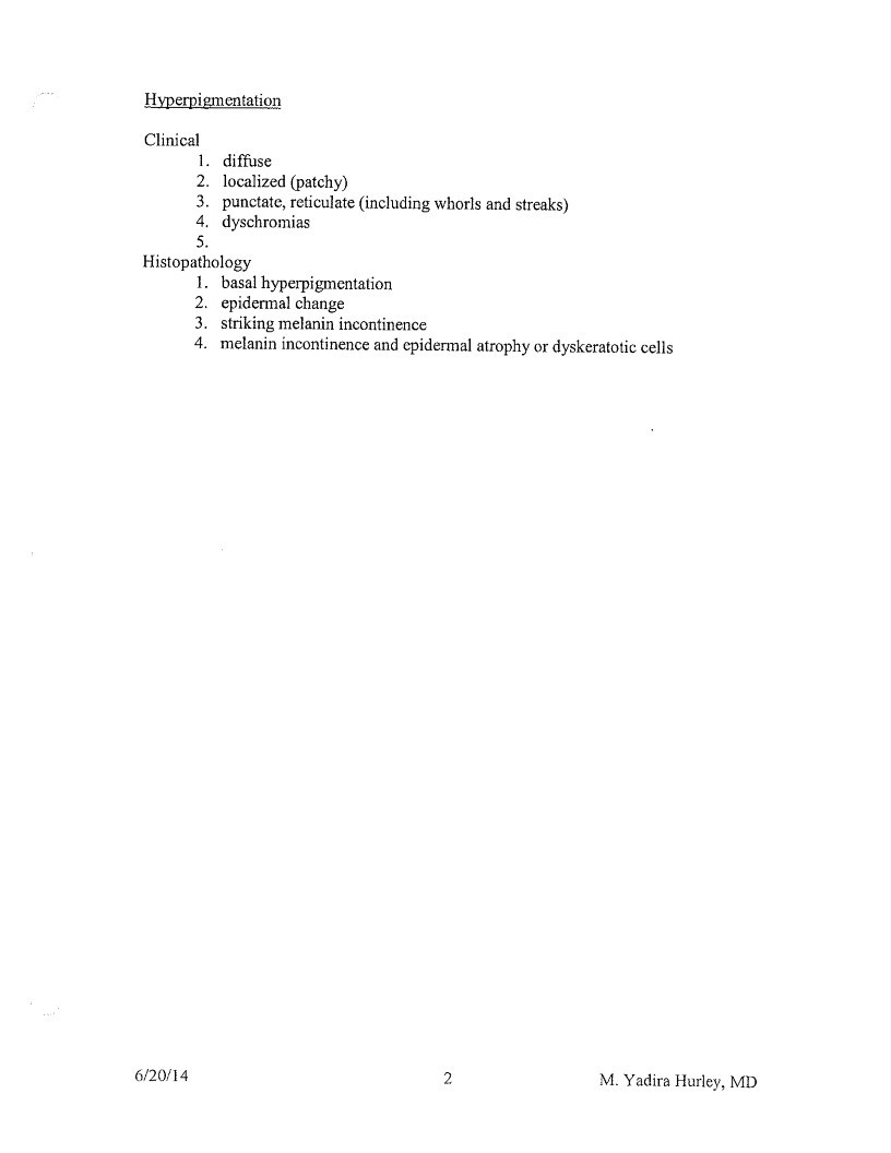
Hyperpigmentation
Clinical
1.
diffuse
2. localized (patchy)
3. punctate, reticulate (including whorls and streaks)
4. dyschromias
5.
Histopathology
- basal hyperpigmentation
- epidermal change
- striking melanin incontinence
melanin incontinence and epidermal atrophy or dyskeratotic cells
6/20/14
2
M. Yadira Hurley, MD
Page 32
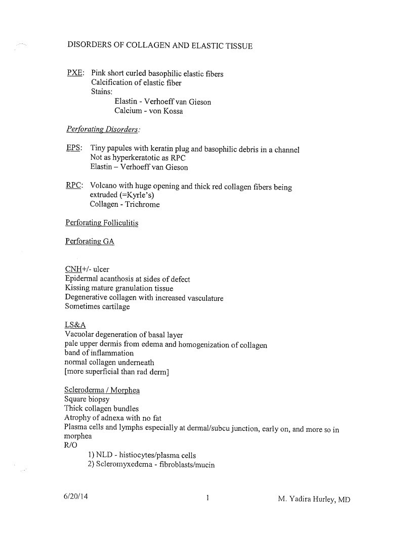
DISORDERS OF COLLAGEN AND ELASTIC TISSUE
PXE: Pink short curled basophilic elastic fibers
Calcification of elastic fiber
Stains:
Elastin - Verhoeff van Gieson
Calcium - von Kossa
Perforating Disorders:
EPS: Tiny papules with keratin plug and basophilic debris in a channel
Not as hyperkeratotic as RPC
Elastin - Verhoeff van Gieson
RPC: Volcano with huge opening and thick red collagen fibers being
extruded (=Kyrle’s)
Collagen - Trichrome
Perforating Folliculitis
Perforating GA
CNH+/- ulcer
Epidermal acanthosis at sides of defect
Kissing mature granulation tissue
Degenerative collagen with increased vasculature
Sometimes cartilage
LS&A
Vacuolar degeneration of basal layer
pale upper dermis from edema and homogenization of collagen
band of inflammation
normal collagen underneath
[more superficial than rad derm]
Scleroderma / Morphea
Square biopsy
Thick collagen bundles
Atrophy of adnexa with no fat
Plasma cells and lymphs especially at dermal/subcu junction, carly on, and more so in
morphea
R/O
- NLD - histiocytes/plasma cells
- Scleromyxedema - fibroblasts/mucin
6/20/14
1
M. Yadira Hurley, MD
Page 33
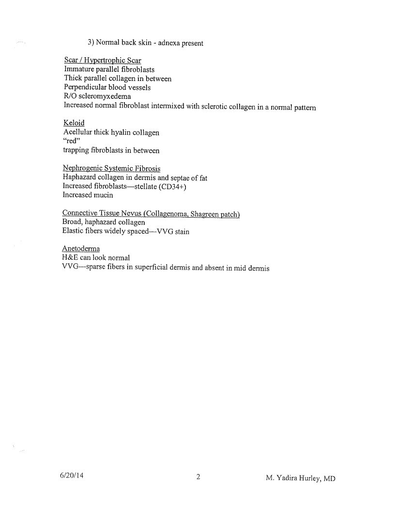
3) Normal back skin - adnexa present
Scar / Hypertrophic Scar
Immature parallel fibroblasts
Thick parallel collagen in between
Perpendicular blood vessels
R/O scleromyxedema
Increased normal fibroblast intermixed with sclerotic collagen in a normal pattern
Keloid
Acellular thick hyalin collagen
”red”
trapping fibroblasts in between
Nephrogenic Systemic Fibrosis
Haphazard collagen in dermis and septae of fat
Increased fibroblasts-stellate (CD34+)
Increased mucin
Connective Tissue Nevus (Collagenoma, Shagreen patch)
Broad, haphazard collagen
Elastic fibers widely spaced—-VVG stain
Anetoderma
H&E can look normal
VVG-sparse fibers in superficial dermis and absent in mid dermis
6/20/14
2
M. Yadira Hurley, MD
Page 34
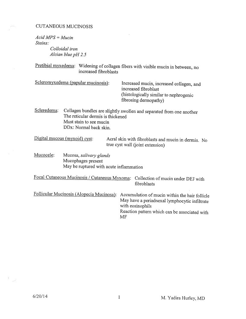
CUTANEOUS MUCINOSIS
Acid MPS = Mucin
Stains:
Colloidal iron
Alcian blue pH 2.5
Pretibial myxedema: Widening of collagen fibers with visible mucin in between, no
increased fibroblasts
Scleromyxedema (papular mucinosis):
Increased mucin, increased collagen, and
increased fibroblast
histologically similar to nephrogenic
fibrosing dermopathy)
Scleredema:
Collagen bundles are slightly swollen and separated from one another
The reticular dermis is thickened
Must stain to sce mucin
DDx: Normal back skin.
Digital mucous (myxoid) cyst:
Acral skin with fibroblasts and mucin in dermis. N
rue cyst wall (joint extensior
Mucocele:
Mucosa, salivary glands
Mucophages present
May be ruptured with acute inflammation
Focal Cutaneous Mucinosis / Cutaneous Myxoma: Collection of mucin under DEJ with
fibroblasts
Follicular Mucinosis (Alopecia Mucinosa): Accumulation of mucin within the hair follicle
May have a periadnexal lymphocytic infiltrate
vith eosinophils
Reaction pattern which can be associated with
MF
6/20/14
1
M. Yadira Hurley, MD
Page 35
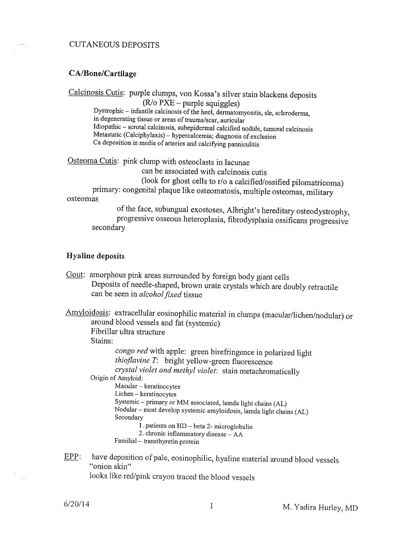
CUTANEOUS DEPOSITS
CA/Bone/Cartilage
Calcinosis Cutis: purple clumps, von Kossa’s silver stain blackens deposits
(R/o PXE - purple squiggles)
Dystrophic - infantile calcinosis of the heel, dermatomyositis, sle, scleroderma,
in degenerating tissue or areas of trauma/scar, auricular
Idiopathic - scrotal calcinosis, subepidermal calcified nodule, tumoral calcinosis
Metastatic (Calciphylaxis) - hypercalcemia; diagnosis of exclusion
Ca deposition in media of arteries and calcifying panniculitis
Osteoma Cutis: pink clump with osteoclasts in lacunae
can be associated with calcinosis cutis
(look for ghost cells to r/o a calcified/ossified pilomatricoma)
primary: congenital plaque like osteomatosis, multiple osteomas, military
osteomas
of the face, subungual exostoses, Albright’s hereditary osteodystrophy,
progressive osseous heteroplasia, fibrodysplasia ossificans progressive
secondary
Hyaline deposits
Gout: amorphous pink areas surrounded by foreign body giant cells
Deposits of needle-shaped, brown urate crystals which are doubly retractile
can be seen in alcohol fixed tissue
Amyloidosis: extracellular eosinophilic material in clumps (macular/lichen/nodular) or
around blood vessels and fat (systemic)
Fibrillar ultra structure
Stains:
EPP:
congo red with apple: green birefringence in polarized light
thioflavine T: bright yellow-green fluorescence
crystai violet and methyl violet: stain metachromatically
Origin of Amyloid:
Macular - keratinocytes
Lichen - keratinocytes
stemic - primary or MIM associated, lamda light chains (A
dular - most develop systemic amyloidosis, lamda light chains (A
Secondary
patients on HD - beta 2- microglobuli
• chronic inflammatory disease - M
Familial - transthyretin protein
have deposition of pale, eosinophilic, hyaline material around blood vessels
”onion skin”
looks like red/pink crayon traced the blood vessels
6/20/14
1
M. Yadira Hurley, MD
Page 36
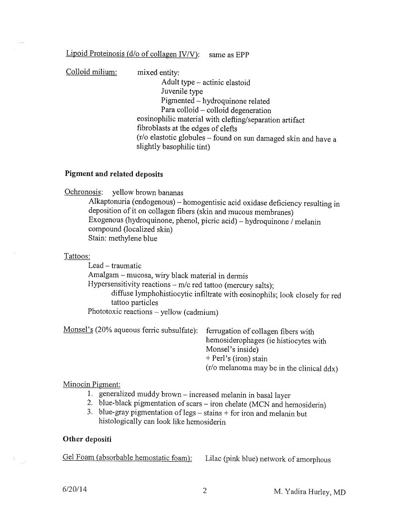
Lipoid Proteinosis (d/o of collagen IV/V):
same as EPP
Colloid milium:
mixed entity:
Adult type - actinic elastoid
Juvenile type
Pigmented - hydroquinone related
Para colloid - colloid degeneration
eosinophilic material with clefling/separation artifact
fibroblasts at the edges of clefts
(r/o elastotic globules - found on sun damaged skin and have a
slightly basophilic tint)
Pigment and related deposits
Ochronosis:
yellow brown bananas
Alkaptonuria (endogenous) - homogentisic acid oxidase deficiency resulting in
deposition of it on collagen fibers (skin and mucous membranes)
Exogenous (hydroquinone, phenol, picric acid) - hydroquinone / melanin
compound (localized skin)
Stain: methylene blue
Tattoos:
Lead - traumatic
Amalgam - mucosa, wiry black material in dermis
Hypersensitivity reactions - m/c red tattoo (mercury salts);
diffuse lymphohistiocytic infiltrate with eosinophils; look closely for red
tattoo particles
Phototoxic reactions - yellow (cadmium)
Monsel’s (20% aqueous ferric subsulfate):
ferrugation of collagen fibers with
hemosiderophages (ie histiocytes with
Monsel’s inside)
- Perl’s (iron) stain
(r/o melanoma may be in the clinical ddx)
Minocin Pigment:
- generalized muddy brown - increased melanin in basal layer
- blue-black pigmentation of scars - iron chelate (MCN and hemosiderin)
- blue-gray pigmentation of legs - stains + for iron and melanin but
histologically can look like hemosiderin
Other depositi
Gel Foam (absorbable hemostatic foam):
Lilac (pink blue) network of amorphous
6/20/14
2
M. Yadira Hurley, MD
Page 37
material in dermis
Other pigment deposits to know:
Blue-Gray with accentuation on sun exposed areas:
Argyria - silver, deposits in basement membrane of eccrine coil
Chrysiasis - gold, deposits around blood vessels, orange-red birefringence
Bismuth - “prurigo pigmentosa”
Phenothiazines - + melanin (female)
Amiodarone - Lipofuscin, + melanin and + Sudan black (male)
Blue-Black - ochronosis
Red-Blue skin and conjunctiva
Clofazimine - Frozen shows birefringent red crystals
Slate Gray
Mercury - large black aggregates in macrophages, around blood vessels, along
elastic fibers
Pretibial/Palate/Subangal
Antimalarials - + hemosiderin and + melanin
Arsenic - normal “rain drops”
Melanin and arsenic deposit in dermis
Yellow
Quinacrine
Blue
Tetracycline - pigmentation of cutaneous osteomas
Blue line on Gingiva
6/20/14
3
M. Yadira Hurley, MD
Page 38
DISEASES OF CUTANEOUS APPENDAGES
Perforating Folliculitis
Folliculitis with rupture of follicle into the dermis
Trichotillomania/Traction Alopecia
Melanin casts within dilated hair follicles
Follicular units without shafts
Alopecia Areata
Peribulbar lymphocytes
Look in subcutancous tissue for anagen hairs
’swarm of bees’
Acne Keloidalis
Naked hair shafts within dense inflammation
Scar/keloid formation
Pseudopelade
Very few hair follicles and no inflammation on scalp
End stage alopecia
Eosinophilic folliculitis
Folliculitis with mostly eosinophils
6/20/2014
1
M. Yadira Hurley, MD
Page 39
CYSTS AND SINUSES
Keratinous Cyst, Infundibular Type
From infundibular portion of follicle
Like skin
Granular layer
Flaky keratin
Keratinous Cyst, Trichilemmal Type
From bottom portion of hair follicle
Like hair
No granular layer
Compact keratin
M/C on scalp
Proliferating Trichilemmal Cyst
KCT with proliferation of cyst wall
Thought to be “SCC” therefore complete excision is required
Older women on back of head
Vellus Hair Cyst
Small, multiple vellus hairs in center
Steatocystoma
Cyst with red cuticle
Sebaceous glands in wall
May be difficult to find
+/- Vellus hairs
Types:
Solitary
Multiplex
Associated with PC type 2
Ecerine Hydrocystoma
Simple cyst
Around eye - look for vellus hairs
2 cell layers:
Myoepithelial
Cuboidal
Apocrine Cystadenoma = Apocrine Hydrocystoma
Truly an adenoma
Occurs anywhere on body
Cyst with multiple invaginations
Cuboidal cells with decapitation secretion
Bronchogenic Cyst
Pseudostratified columnar ciliated lining
+/- goblet cells (blue)
6/20/14
1
M. Yadira Hurley, MD
Page 40
Dermoid Cyst
Hamartoma:
Eccrine glands
Apocrine glands
Pilosebaceous units
If you have a cyst check the wall carefully for eccrine and apocrine glands and pilosebaceous
units
Found at embryonic fusion lines
Pseudocyst of the Auricle
Not a true cyst because it has no lining
With in cartilage of ear
Can reoccur
Endometriosis
Fibrotic stroma
Glandular structures with central RBC in dermis
6/20/14
2
M. Yadira Hurley, MD
Page 41
PANNICULITIS
Septal
- Erythema nodosum
- NLD
- Scleroderma
Lobular - Nodular vasculitis
- Subcutaneous Fat Necrosis
- Alpha one antitrypsin deficiency
- Cytophagic histiocytic panniculitis = lymphoma
- Pancreatic panniculitis
- Lupus panniculitis
- Lipodermatosclerosis
- Sclerosing Lipogranuloma
SEPTAL
Erythema Nodosum
Erythema Nodosum: Widened fibrous septae
Giant cells in septae = Miescher’s granulomas
May/may not have eos, neuts
No vasculitis or necrosis
LOBULAR
Nodular Vasculitis
= Erythema Induratum
”Vasculitis” at dermal-sc junction and in septa (small and medium vessels)
Lobules with neutrophils-> fat necrosis => foamy macrophages
Involves contiguous lobules
R/O infection
vS. PAN - large vessels in septa, restricted panniculitis
Subcutaneous Fat Necrosis of the Newborn - Sclerema Neonatorum
Normal Dermis
Needle like clefts in lipocytes
+/- Giant cells in fat
Pancreatic Panniculitis
Focal areas in center of lobule
Coagulative necrosis of lipocytes =>pink no nucleus
calcium deposits on lipocytes = ghost cells => blue rim
6/20/14
1
M. Yadira Hurley, MD
Page 42
Surrounded by neutrophils
Pink - Blue - PMNs
Lupus Panniculitis
Dense lymphoplasmacytic infiltrate (swiss cheese lobules)
Lymphoid follicles at edges of fat lobules
Nuclear debris from lymphs
+/- lupus changes on epidermis and dermis (50%)
- lupus band on DIF
/o subcutaneous panniculitis like T-cell lymphoma
Lipodermatosclerosis
= Membranous lipodystrophy / Sclerosing Panniculitis
Stasis changes at the top
Thick sclerosed septa
Cystic degencration of fat = large holes
Lipo membranous changes = feathery cuticle inside cysts
Sclerosing Lipogranuloma
= paraffinoma
No dermis or epidermis seen
Disruption of fat cells with replacement by cystic spaces of variable size
Cyst in fibrous tissue
Chunk of deep tissue with swiss cheese look
Alpha 1 antitrypsin Deficiency
Pouring of neutrophils into reticular dermis and SQ septae
Splaying of neutrophils between collagen bundles
Dissolution of septae and dermis
6/20/14
2
M. Yadira Hurley, MD
Page 43
METABOLIC AND STORAGE DISEASE / MISCELLANEOUS
rabry s Disease (Angiokeratoma corporis diffusum)
See angiokeratomas on routine histology (Vascular tumors chapter)
EM-Intracytoplasmic electron-dense lamellar inclusion bodies within endothelial cells
Necrolytic Migratory Erythema (Glucagonoma)
Acrodermatitis Enteropathica
Both have identical path
Three layer epidermis:
- Parakeratosis / Hyperkeratosis (pink)
- Pale vacuolated keratinocytes (pale)
- Normal epidermis (normal)-may have psoriasiform hyperplasia
Diabetic Dermopathy
Looks similar to stasis changes
Blood vessel proliferation in superficial dermis with perivascular lymphocytes and
hemosiderophages.
PCI
Subepidermal blister with few lymphoevtes
”Caterpillar bodies:—elongated bodies or globules of basement membrane material and
colloid bodies in the basal layer of the epidermis
Thick BMZ around vessels-PAS positive and diastase resistant
Festooning (more common b/c of thick vessels with BMZ but any subepidermal blister
can have this)
Look for solar clastosis to differentiate from EPP
DIF-IgG around upper dermal blood vessels
Accessory Tragus
+/- Cartilage in center
Polypoid piece of tissue
Central fat
Rim of vellus hair
Preauricular
Supernumerary Nipple
Epidermal thickening w/ mild papillomatosis and basal hyperpigmentation
Central invaginated enlarged pilosebaceous unit
Smooth muscle hyperplasia in the dermis
Modified, deep, dark staining apocrine glands / ducts
Trichrome stain-muscle red
Smooth muscle in skin:
Scrotum
Pilar - leiomyoma
6/20/14
1
M. Yadira Hurley, MD
Page 44
Arteries - angioleiomyoma
Relapsing Polychondritis
Degeneration of cartilage with florid perichondritis (neutrophils initially with
progressively more lymphocytes, plasma cells and histiocytes)
Type II collagen
Acanthosis Nigricans
Hyperkeratosis, papillomatosis and mild acanthosis
Basal keratinocyte pigmentation
No inflammation
Histology very similar to:
Subtype of epidermal nevus
Becker’s melanosis (nevus)
Associations: Insulin resistance and “malignancy”
Confluent and Reticulated Papillomatosis
Undulating epidermis with hyperkeratosis and low papillomatosis
Mild basal hyperpigmentation
Resembles AN but not as well developed
6/20/14
2
M. Yadira Hurley, MD
Page 45
REACTIONS TO PHYSICAL AGENTS
PMLE
Marked subepidermal edema
Superficial and deep perivascular lymphocytic infiltrate
Photodermatosis
• Phototoxic
Clinically = sunburn
Histology: Sun burn cells = apoptotic keratinocytes
Usually orals
Dose dependant
Photoallergic
Histology = allergic contact dermatitis
Usually topicals
Except for thiazides
May persist despite removal of allergen
Radiation Dermatitis
Epidermal atrophy
Telangiectatic blood vessel
Loss of adnexa
Hyalinized collagen - pink
Stellate fibroblasts
Bur
Electrical = Thermal
Epidermis with elongated keratinocytes
Homogeneous collagen
Freeze artifact - perinuclear bubble, pink
Air dry artifact - cells with clear center in epidermis
Crush Artifact
m/c in hematopoietic infiltrates
6/20/14
Yadira Hurley, MD
Page 46
BACTERIAL INFECTIONS
- Bullous impetigo
-Subcorneal cleft with acantholysis, neutrophils and gram positive cocci
-Mild to moderate mixed infiltrate - Staphylococcal scalded skin syndrome (SSSS)
-Subcorneal cleft with acantholysis, sparse neutrophils and no bacteria
-Sparse mixed infiltrate - Erythrasma
- “Normal skin” differential
-Small coccobacilli in stratum corneum on Gram stain
- Botryomycosis
-Basophilic granule (bacteria) surrounded by eosinophilic material (Splendore-
Hoeppli phenomenon) within an area of suppuration - Atypical mycobacterial infection
-acute abscesses, suppurative granulomas or poorly formed granulomas
-organısms stain with acid fast stain (red snappers) -look for them in cystic spaces - Leprosy
- Lepromatous leprosy: sheets of heavily parasitized macrophages and can look
Foamy (lepra or Virchow cells); clumps of acid-fast bacilli in macs (globi)-.
Fite stain (acid-fast variant) may highlight them better
-Tuberculoid leprosy: non-cascating granulomas with epithelioid cells, Langhans giant
cells and lymphocytes; bacilli are rare
-Indeterminate leprosy: superficial and deep dermal infiltrate of lymphocytes and few
histiocytes around blood vessels, appendages and nerves; can usually find few
AFB
- Rhinoscleroma
-Sheets of vacuolated macrophages (Mikulicz cells) containing Gram negative bacilli
best seen with Warthin-Starry
-Plasma cells with prominent Russell bodies (aggregates of immunoglobulin) - Secondary syphilis
- Variable histology
-Lichenoid reaction pattern with plasma cells and endothelial cell swelling is classic
- Bacillary angiomatosis
-Closely resembles a pyogenic granuloma with a lobular proliferation of blood vesseis;
however neutrophils are much more prominent in BA
-Plump endothelial cells
-Clumps of granular material (bacteria) -seen on GMS or Warthin-Starry
Page 47
FUNGAL INFECTIONS
- Tinea corporis
-Normal skin differential
-Stratum corneum changes: neutrophils, compact orthokeratosis or the “sandwich
sign” (hyphae in between upper normal basket weave SC and lower
compact orthokeratotic or parakeratotic scale)
-Can have mild spongiosis and acanthosis
-Lymphocytic infiltrate +/- eos/neuts - Majocchi’s granuloma
-Perifollicular and dermal granulomas and mixed acute and chronic inflammation
-Fungal elements in follicle and in the dermis - Candidiasis
-Neutrophils in the stratum corneum
-Epithelium may demonstrate mild spongiosis and acanthosis
-Budding fungal yeast forms - Tinea versicolor
-Normal skin differential
-Ycast forms and short hyphae in stratum corneum-can see well on H&E - Cryptococcosis
-Epidermis may show ulceration or acanthosis
-Acute and chronic inflammation w/ MNGCs
-Can have a mucinous appearance due to the mucinous capsular material
-Encapsulated yeast-like organisms
-Cell wall stains with PAS, GMS or Fontana-Masson (also a silver stain)
-Capsule stains w/ mucicarmine or Alcian blue - Blastomycosis
-Epidermis can have pseudoepitheliomatous hyperplasia (CBS—cocci, blasto,
sporo are classic)
-Suppurative granulomatous inflammation
-Within abscesses and in giant cells can see thick-walled yeasts w/ broad-based
budding - Phacohyphomycosis
-Circumscribed cyst or abscess in the deep dermis/subcutis
-Brown filamentous hyphae and yeast forms present (as opposed to
chromoblastomycosis in which brown, round sclerotic or medlar bodies
are seen - Mucormycosis
-Suppuration or areas of necrosis
Page 48
-Broad, non-septate hyphae in blood vessel walls seen casily on H&E (branch at
90 degrees)
VIRAL INFECTIONS
- Molluscum contagiosum
-Inverted lobules of hyperplastic squamous epithelium
-Eosinophilic inclusion bodies (Henderson-Patterson bodies) within the
keratinocyte cytoplasm - Herpesvirus infections
-Histology of herpes simples, varicella and herpes zoster are very similar
-Keratinocytes develop peripheral clumping (margination) of chromatin,
homogenous ground-glass appearance, vacuolization of cytoplasm, and
eosinophilic intranuclear inclusion bodies
-Intrepidermal vesicle forms from two types of degenerative change:
a. Ballooning degeneration: affected keratinocytes swell and lose
attachment to adjacent cells (acantholysis)
b. Reticular degeneration: Progressive swelling of keratinocytes w/
only tine cytoplasmic strands
-Multinucleated keratinocytes may also be present
-Can involve pilosebaceous units
-Vasculitis more common in zoster - Cytomegalovirus
- Enlarged endothelial cells with intranuclear eosinophilic inclusions surrounded
by a halo (owl’s eye)
- Verruca vulgaris
-Inward turning of elongated rete ridges
-Columns of parakeratosis over papillomatous projections
- Clumping of keratohyaline granules
-Koilocytes: in superficial cpidermis; small pyknotic nucleus surrounded by clear
cytoplasm
- Myrmecial wart
- “Myrmecia”=anthill
-Deep extensions of acanthotic epidermis
-Large cosinophilic cytoplasmic inclusions
- Verruca plana
-Basket-weave hyperkeratosis and mild acanthosis
-Vacuolation of upper keratinocytes - Condyloma acuminatum
-Marked acanthosis with broad rete, hyperkeratosis and parakeratosis
Page 49
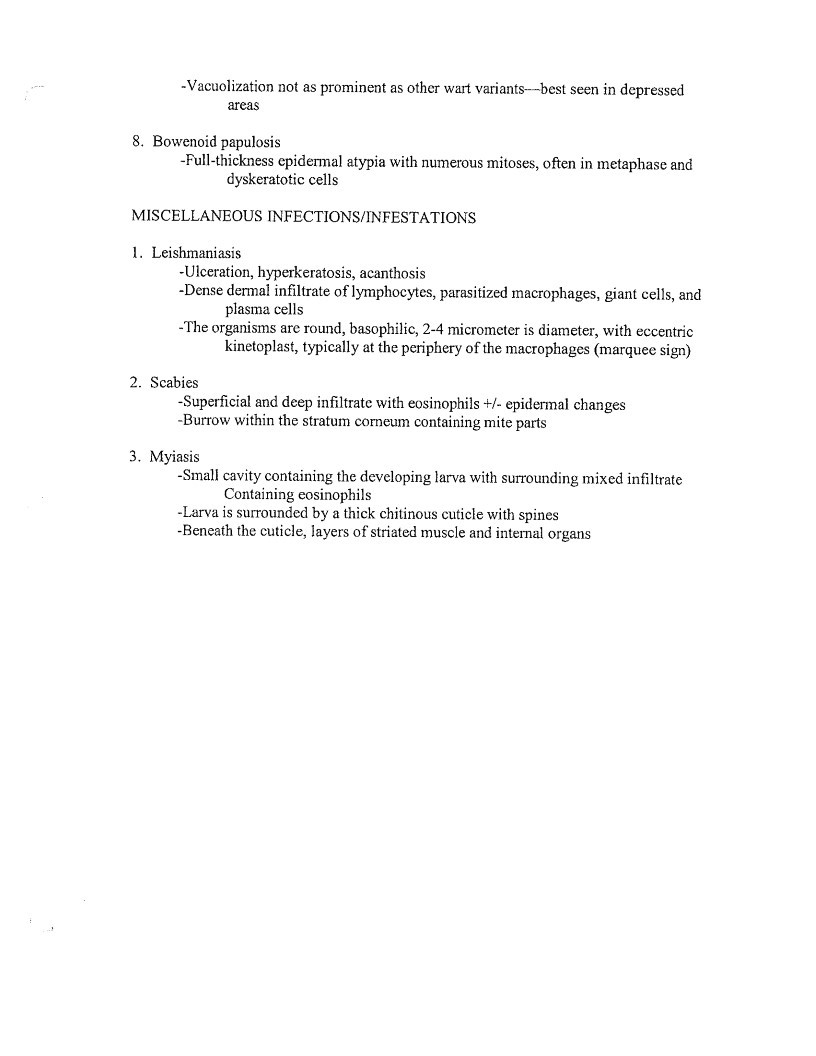
-Vacuolization not as prominent as other wart variants-—-best seen in depressed
areas
8. Bowenoid papulosis
-Full-thickness epidermal atypia with numerous mitoses, often in metaphase and
dyskeratotic cells
MISCELLANEOUS INFECTIONS/INFESTATIONS
- Leishmaniasis
-Ulceration, hyperkeratosis, acanthosis
-Dense dermal infiltrate of lymphocytes, parasitized macrophages, giant cells, and
plasma cells
-The organisms are round, basophilic, 2-4 micrometer is diameter, with eccentric
kinetoplast, typically at the periphery of the macrophages (marquee sign) - Scabies
-Superficial and deep infiltrate with eosinophils +/- epidermal changes
- Burrow within the stratum corneum containing mite parts
- Myiasis
-Small cavity containing the developing larva with surrounding mixed infiltrate
Containing eosinophils
- Larva is surrounded by a thick chitinous cuticle with spines
-Beneath the cuticle, layers of striated muscle and internal organs
Page 50
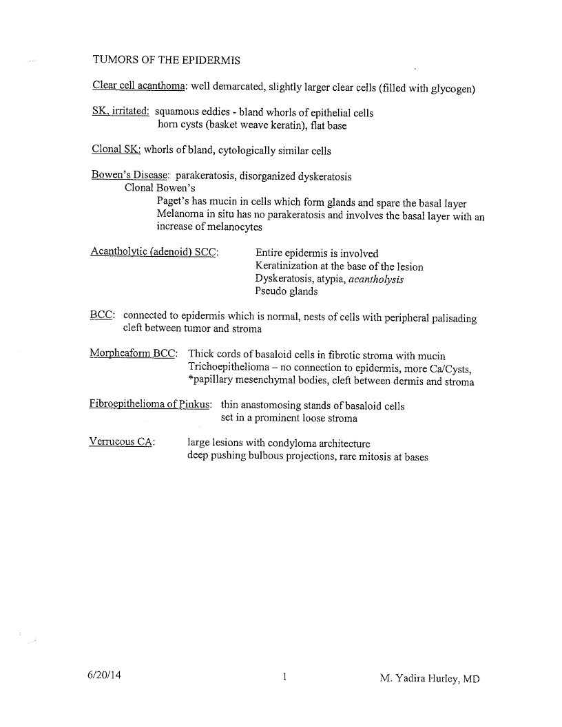
TUMORS OF THE EPIDERMIS
Clear cell acanthoma: well demarcated, slightiy larger clear cells (filled with glycogen)
SK, irritated: squamous eddies - bland whorls of epithelial cells
horn cysts (basket weave keratin), flat base
Clonal SK: whorls of bland, cytologically similar cells
Bowen’s Discase: parakeratosis, disorganized dyskeratosis
Clonal Bowen’s
Paget’s has mucin in cells which form glands and spare the basal layer
Melanoma in situ has no parakeratosis and involves the basal layer with an
increase of melanocytes
Acantholytic (adenoid) SCC:
Entire epidermis is involved
Keratinization at the base of the lesion
Dyskeratosis, atypia, acantholysis
Pseudo glands
BCC: connected to epidermis which is normal, nests of cells with peripheral palisading
cleft between tumor and stroma
Morpheatorm BCC: Thick cords of basaloid cells in fibrotic stroma with mucin
Trichoepithelioma - no connection to epidermis, more Ca/Cysts,
*papillary mesenchymal bodies, cleft between dermis and stroma
Fibroepithelioma of Pinkus: thin anastomosing stands of basaloid cells
set in a prominent loose stroma
Verrucous CA:
large lesions with condyloma architecture
deep pushing bulbous projections, rare mitosis at bases
6/20/14
1
M. Yadira Hurley, MD
Page 51
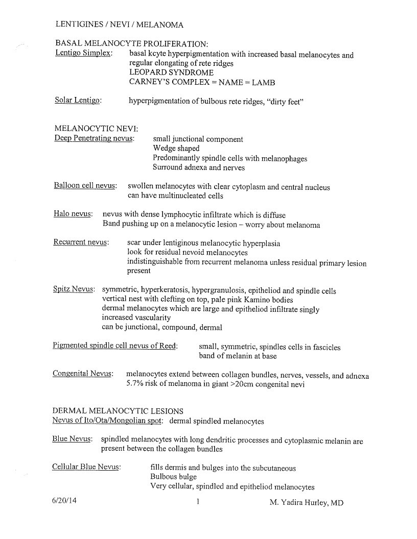
LENTIGINES / NEVI / MELANOMA
BASAL MELANOCYTE PROLIFERATION:
Lentigo Simplex:
basal kcyte hyperpigmentation with increased basal melanocytes and
regular clongating of rete ridges
LEOPARD SYNDROME
CARNEY’S COMPLEX = NAME = LAMB
Solar Lentigo:
hyperpigmentation of bulbous rete ridges, “dirty feet”
MELANOCYTIC NEVI:
Deep Penetrating nevus:
small junctional component
Wedge shaped
Predominantiy spindle cells with melanophages
Surround adnexa and nerves
Balloon cell nevus:
wollen melanocytes with clear cytoplasm and central nucleu
an have multinucleated cell
Halo nevus:
Recurrent nevus:
nevus with dense lymphocytic infiltrate which is diffuse
Band pushing up on a melanocytic lesion - worry about melanoma
scar under lentiginous melanocytic hyperplasia
look for residual nevoid melanocytes
indistinguishable from recurrent melanoma unless residual primary lesion
present
Spitz Nevus:
symmetric, hyperkeratosis, hypergranulosis, cpitheliod and spindle cells
vertical nest with clefting on top, pale pink Kamino bodies
dermal melanocytes which are large and epitheliod infiltrate singly
increased vascularity
can be junctional, compound, dermal
Pigmented spindle cell nevus of Reed:
small, symmetric, spindles cells in fascicles
band of melanin at base
Congenital Nevus:
melanocytes extend between collagen bundles, nerves, vessels, and adnexa
5.7% risk of melanoma in giant >20cm congenital nevi
DERMAL MELANOCYTIC LESIONS
Nevus of Ito/Ota/Mongolian spot: dermal spindled melanocytes
Blue Nevus: spindled melanocytes with long dendritic processes and cytoplasmic melanin are
present between the collagen bundles
Cellular Blue Nevus:
fills dermis and bulges into the subcutaneous
Bulbous bulge
Very cellular, spindled and epitheliod melanocytes
6/20/14
M. Yadira Hurley, MD
Page 52
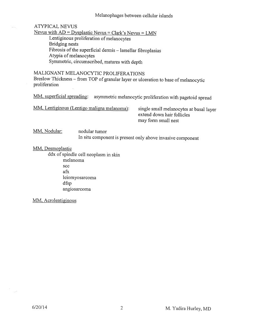
Melanophages between cellular islands
ATYPICAL NEVUS
Nevus with AD = Dysplastic Nevus = Clark’s Nevus = LMN
Lentiginous proliferation of melanocytes
Bridging nests
Fibrosis of the superficial dermis - lamellar fibroplasias
Atypia of melanocytes
Symmetric, circumscribed, matures with depth
MALIGNANT MELANOCYTIC PROLIFERATIONS
Breslow Thickness - from TOP of granular layer or ulceration to base of melanocytic
proliferation
MM, superficial spreading:
asymmetric melanocytic proliferation with pagetoid spread
MM, Lentiginous (Lentigo maligna melanoma):
single small melanocytes at basal layer
extend down hair follicles
may form small nest
MM, Nodular:
nodular tumor
In situ component is present only above invasive component
MM, Desmoplastic
ddx of spindle cell neoplasm in skin
melanoma
SCC
afx
leiomyosarcoma
disp
angiosarcoma
MM. Acrolentiginous
6/20/14
2
M. Yadira Hurley, MD
Page 53
TUMORS OF CUTANEOUS APPENDAGES
HAIR FOLLICLE TUMORS
HAMARTOMAS
Trichofolliculoma
Small follicles with variable maturity radiate from large central
follicle
Trichocpithelioma
Multiple nest of basaloid cells or interlacing pattern, symmetric,
3 connect to epidermis, cleft between stroma and epidermis, ca
ave cysts, look for abortive papillary mesenchymal bodie
Dax: bcc
Desmoplastic Trichoepithelioma
linear cords and islands of basaloid cells in fibrous stroma,
keratinous cysts, FBGCR, and calcium
Ddx: morpheaform bec
INFUNDIBULAR / ISTHMUS TUMORS
Dilated Pore of Winer
Finger like projections from a dilated follicular pore
Inverted follicular keratosis
Endophytic tumor, basaloid cells at the periphery with larger
keratinizing cells in the center
Look for squamous eddies at base
EXTERNAL SHEATH TUMORS
Trichilemmoma
lobular vertical growth, glycogen vacuolation of squamoid cells,
erpheral layer of columnar cells with palisading, thick basemen
membrane surrounds tumor; PAS-
*Cowden’s
MATRICAL TUMORS
Pilomatrixoma
Sharply demarcated tumors in dermis, two types of cells:
basophilic cells at the periphery and eosinophilic shadow cells in
central areas
Can have calcification and ossification
*Multiple - Myotonic Dystrophy
PERIFOLLICULAR MESENCHYME
Fibrofolliculoma / trichodiscoma
Central hair follicle like structure with surrounding well-
circumscribed proliferation of loose connective tissue
*B-H-D
6/20/14
1
M. Yadira Hurley, MD
Page 54
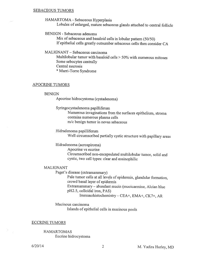
SEBACEOUS TUMORS
HAMARTOMA - Sebaceous Hyperplasia
Lobules of enlarged, mature sebaceous glands attached to central follicle
BENIGN - Sebaceous adenoma
Mix of sebaceous and basaloid cells in lobular pattern (50/50)
If epithelial cells greatly outnumber sebaceous cells then consider CA
MALIGNANT - Sebaceous carcinoma
Multilobular tumor with basaloid cells > 50% with numerous mitoses
Some sebocytes centrally
Central necrosis
- Murri-Torre Syndrome
APOCRINE TUMORS
BENIGN
Apocrine hidrocystoma (cystadenoma)
Syringocystadenoma papilliferum
Numerous invaginations from the surfaces epithelium, stroma
contains numerous plasma cells
m/c benign tumor in nevus sebaceous
Hidradenoma papilliferum
Well circumscribed partially cystic structure with papillary areas
Hidradenoma (acrospiroma)
Apocrine vs eccrine
Circumscribed non-encapsulated multilobular tumor, solid anc
ystic, two cell types: clear and eosinophilic
MALIGNANT
Paget’s disease (extramammary)
Pale tumor cells at all levels of epidermis, glandular formation,
crowd basal layer of epidermis
Extramammary - abundant mucin (mucicarmine, Alcian blue
H2.5, colloidal iron, PAS
Immunohistochemistry - CEA+, EMA+, CK7+, AR
Mucinous carcinoma
Islands of epithelial cells in mucinous pools
ECCRINE TUMORS
HAMARTOMAS
Eccrine hidrocystoma
6/20/14
2
M. Yadira Hurley, MD
Page 55
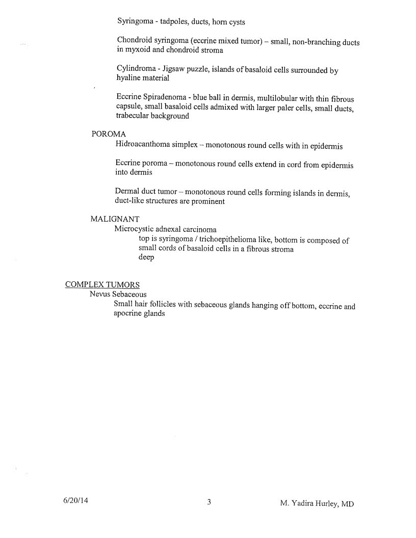
Syringoma - tadpoles, ducts, horn cysts
Chondroid syringoma (eccrine mixed tumor) - small, non-branching ducts
in myxoid and chondroid stroma
Cylindroma - Jigsaw puzzle, islands of basaloid cells surrounded by
hyaline material
Eccrine Spiradenoma - blue ball in dermis, multilobular with thin fibrous
capsule, small basaloid cells admixed with larger paler cells, small ducts,
trabecular background
POROMA
Hidroacanthoma simplex - monotonous round cells with in epidermis
Eccrine poroma - monotonous round cells extend in cord from epidermis
into dermis
Dermal duct tumor - monotonous round cells forming islands in dermis,
duct-like structures are prominent
MALIGNANT
Microcystic adnexal carcinoma
top is syringoma / trichoepithelioma like, bottom is composed of
small cords of basaloid cells in a fibrous stroma
deep
COMPLEX TUMORS
Nevus Sebaceous
Small hair follicles with sebaceous glands hanging off bottom, eccrine and
apocrine glands
6/20/14
3
M. Yadira Hurley, MD
Page 56
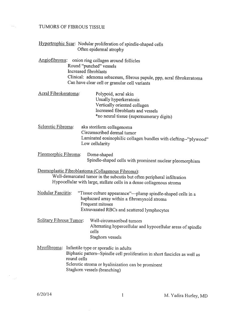
TUMORS OF FIBROUS TISSUE
Hypertrophic Scar: Nodular proliferation of spindle-shaped cells
Often epidermal atrophy
Angiofibroma: onion ring collagen around follicles
Round “punched” vessels
Increased fibroblasts
Clinical: adenoma sebaceum, fibrous papule, ppp, acral fibrokeratoma
Can have clear cell or granular cell variants
Acral Fibrokeratoma:
Sclerotic Fibroma:
Polypoid, acral skin
Usually hyperkeratosis
Vertically oriented collagen
Increased fibroblasts and vessels
*no neural tissue (supernumerary digits)
aka storiform collagenoma
Circumscribed dermal tumor
Laminated eosinophilic collagen bundles with clefting.. “plywood”
Low cellularity
Pleomorphic Fibroma:
Dome-shaped
Spindle-shaped cells with prominent nuclear pleomorphism
Desmoplastic Fibroblastoma (Collagenous Fibroma):
Well-demarcated tumor in the subcutis but often peripheral infiltration
Hypocellular with large, stellate cells in a dense collagenous stroma
Nodular Fasciitis:
“Tissue culture appearance”—-plump spindle-shaped cells in a
haphazard array within a fibromyxoid stroma
Frequent mitoscs
Extravasated RBCs and scattered lymphocytes
Solitary Fibrous Tumor:
Well-circumscribed tumors
Alternating hypercellular and hypocellular areas of spindle
Staghorn vessels
Myofibroma: Infantile type or sporadic in adults
Biphasic pattern—Spindle cell proliferation in short fascicles as well as
round cells
Sclerotic stroma or hyalinization can be prominent
Staghorn vessels (branching)
6/20/14
M. Yadira Hurley, MD
Page 57
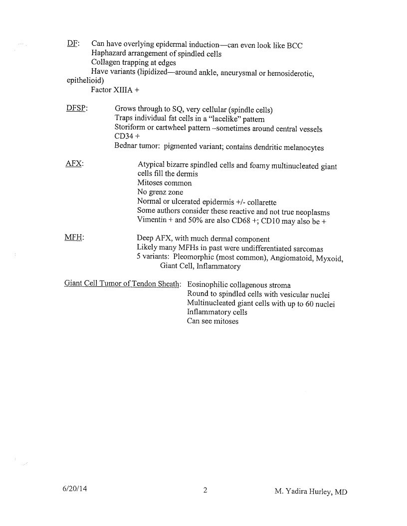
DF:
Can have overlying epidermal induction—-can even look like BCC
Haphazard arrangement of spindled cells
Collagen trapping at edges
Have variants (lipidized—around ankle, aneurysmal or hemosiderotic,
epithelioid)
Factor XIIIA +
DFSP:
Grows through to SQ, very cellular (spindle cells)
Traps individual fat cells in a “lacelike” pattern
Storiform or cartwheel pattern -sometimes around central vessels
CD34 +
Bednar tumor: pigmented variant; contains dendritic melanocytes
APX:
Atypical bizarre spindled cells and foamy multinucleated giant
cells fill the dermis
Mitoses common
No grenz zone
Normal or ulcerated epidermis +/- collarette
Some authors consider these reactive and not true neoplasms
Vimentin + and 50% are also CD68 +; CD10 may also be +
MFH:
Deep AFX, with much dermal component
Likely many MFHs in past were undifferentiated sarcomas
5 variants: Pleomorphic (most common), Angiomatoid, Myxoid,
Giant Cell, Inflammatory
Giant Cel! Tumor of Tendon Sheath: Eosinophilic collagenous stroma
Round to spindled cells with vesicular nuclei
Multinucleated giant cells with up to 60 nuclei
Inflammatory cells
Can see mitoses
6/20/14
M. Yadira Hurley, MD
Page 58
TUMORS OF FAT AND MUSCLE/CARTILAGE/BONE
Angiolipoma: fat and blood vessels with thrombi at periphery
Hibernoma:
“brown fat”
3 cells: large cells with multiple vacuoles (filled with ping-pong balls)
uni vacuolated cells
small cells with granular cytoplasm
Myxoid Liposarcoma:
Network of collapsed vessels - “Chicken wire appearance”
Mucoid stroma
Vacuolated lipoblasts and stellate cells
Very deep tumor
Leiomyoma:
Somewhat circumscribed non encapsulated tumor in dermis
Red-orange bundles of smooth muscle with whorled pattern
Cigar shaped nuclei run in fascicles
Angioleiomyoma:
Deep red-orange nodular tumor
Collapsed vessels with thick walls of smooth muscle that merge
Sometimes no epidermis or dermis present
Leiom yosarcoma:
Spindled shaped cells with cigar-shaped nuclei - prominent mitosis
Nodular or diffuse pattern
6/20/2014
1
Yadira Hurley, MD
Page 59
NEURAL AND NEUROENDOCRINE TUMORS
Rudimentary polydactyly (supernumerary digit/amputation neuroma):
polypoid acral surface
Small “Meissner’s corpuscles”
Solitary circumscribed neuroma (palisaded and encapsulated neuroma):
Located in upper dermis
Nerve fascicles without collagen in between
Well demarcated with clefting
Schwannoma (neurilemmoma):
Circumscribed, encapsulated tumor
Antoni A - cellular, fascicles, Verocay bodies
(picket fence nuclei)
Antoni B - acellular CT
NF:
wavy, delicate spindle-shaped nuclei and pink wispy stroma
+mast cells and small vessels
Granular cell tumor: Polyhedral cells with granular cosinophilic cytoplasm
Neuroendocrine Carcinoma (Merkel cell tumor):
small round blue cells with indistinct cytoplasmic boarders
Architecture: trabecular or grape like clusters
Nuclei: sait and pepper
Stains:
CK20 - paranucicar dot positivity
Chromogranin
Synaptophysin
6/20/14
1
Yadira Hurley, MD
Page 60
VASCULAR TUMORS
Lymphangioma:
Endothelial lining with milky substance (no rbes)
valves
Venous Lake:
Solitary large vessel in upper dermis + thrombus
Angiokeratoma:
Epidermal acanthosis and elongated rete ridges surrounding dilated vascular channels
Types: Mibelli
Fordyce
Solitary and multiple
Circumscriptum
Fabry’s (corporis diffusum) - lamellar lipid bodies
AV Hemangioma: Large thick-walled vessels with fibromuscular wall
Microvenular Hemangioma: Thin uniform branching collapsed vessels
Targetoid Hemosiderotic Hemangioma: Small vessels with hobnail endothelial cells
Rbe and hemosiderin
Dax: Kaposi’s sarcoma
ALHE:
Myxoid and fibrous stroma around vessels with plump endothelial cells (look epithelioid)
Lymphocytes (may form follicles) and eosinophils
Eosinophils on H+N: Granuloma Faciale or ALHE
Kimura’s: eosinophilic lymphogranuloma has same histology
LCH:
Lobular vascular proliferation with well developed collarette of epidermis
Fibrous septae separate lobules
No layering of roc r/o Kaposi’s
May have pmns when ulcerates r/o bacillary angiomatosis
Bacillary angiomatosis:
Looks like loch with pmns but no ulceration
Organisms make purple granular clumps
Bartonella hensela quintana
Glomus tumor:
Rounded monotonous cells with eosinophilic cytoplasm and dark round nuclei
Several layers thick around vessels - Glomangioma
Mostly cells - Glomus Tumor
Intravascular papillary endothelial hyperplasia: (Masson’s tumor, organizing hematoma)
Vascular channels within collagenous thrombus surrounded by vessel wall
Papillary fronds covered by endothelial cells
Kaposi’s sarcoma:
Irregular jagged slit like vascular spaces
Promontory sign: slit like vascular space surround normal preexisting blood vessel
RBCs in parallel trapped by spindled cells (layered look)
Plasma cells
Eosinophilic globules
Hemosiderin especially early
6/20/14
Yadira Hurley, MD
Page 61
Angiosarcoma:
Anastomosing, clefting vessels that are dissecting (“raining”) down
Highly atypical endothelial cells: spindled to polygonal
Endothelial cells crowd the vessels and form papillary collections
In differential of malignant spindle cell tumor of dermis
Granulation tissue:
Edematous background
Vessels engorged by rbo
Neut, plasma, lymphs, histiocytes
6/20/14
2
Yadira Hurley, MD
Page 62
CUTANEOUS METS
Renal Cancer Met
Dermal tumor composed of clear cells with prominent vascularity
Common on scalp
Breast Cancer Met
Linear cords of cells that touch and can form glandular structures
”Indian file”
Usually less cellular than leukemia cutis
Malignant Melanoma Met
Dermal tumor with nesting and focal areas of pigment
Frequent mitoses
6/20/2014
1
M. Yadira Huriey, MD
Page 63
CUTANEOUS INFILTRATES
Mastocytosis
Mononuclear cell infiltrate in dermis
Usually eosinophils also present
Cells have granular cytoplasm
LCH
Epidermotropism of large cells
Cells are large with kidney shaped pale nuclei
Dermal edema with cells floating
Can have eosinophils associated
JXG
Touton giant cells (wreath with surrounding foam)
Dermal tumor of mononuclear cells
Eosinophils and lymphocytes also present
Xanthelasma
Thin epidermis with many small hair follicles
Mononuclear cells with foamy cytoplasm/lipid in all cells
Lipid can be extracellular
Reticulohistiocytic Granuloma
Giant cells with pink ground glass cytoplasm
6/20/2014
1
M. Yadira Hurley, MD
Page 64
LYMPHOID AND LEUKEMIC INFILTRATES
MF
Most common type of cutaneous lymphoma overall (T-cell and B-cell)
Subtype of Cutaneous T-cell lymphoma
Collections of lymphocytes in cidermis with minimal spongiosis
= Pautrier microabscesses
Lymphocytes line up at the DEJ but do not obscure
’pearl necklace’
Lymphocytes are larger, have a cerebriform nuclei and a clear perinuclear halo
CD3+/CD4+ / CD7 decreased /CD8-/CD30-
Clonal aß TCR gene rearrangements
Classic type patch to plaque to tumor (Alibert-Bazin type)
Other subtypes of CTCL with distinctive histologic and clinical findings:
Folliculotropism mycosis fungoides, pagetoid reticulosis (Woringer-Kolopp disease),
granulomatous slack skin, Sézary syndrome
LYP (type A)
Wedge shaped infiltrate with cosinophils, neutrophils, large mononuclear cells
Large mononuclear cells have anaplastic morphology (bizarre, chunks of coal)
CD3+/CD4+/CD30+
r/o PLEVA - look at sides for dyskeratotic keratinocytes
r/o lymphoma (primary cutaneous anaplastic large cell lymphoma = C-ALCL)
which would fill dermis and not have a mixed infiltrate
C-ALCL is ALK t (2; 5) negative and epithelial membrane antigen
negative; these are markers pr primary systemic anaplastic large cell
lymphoma
*One of the primary cutaneous CD30+ lymphoproliferative disorder:
• Common histologic feature: large atypical lymphoid cells CD30+
• CD30 is a transmembrane cytokine receptor to TNF receptor
family and is expressed by activated lymphocytes
• Includes: primary cutaneous anaplastic large cell lymphoma,
lymphomatoid papulosis, and borderline cases
- Distinction is based on clinical presentation and course not
possible on histology alone
Cutaneous Lymphoid Hyperplasia
*Pseudolymphoma / lymphadenosis benigna cutis / lymphocytoma cutis /
cutaneous lymphoplasia’
Classically simulates B-cell lymphoma
In Europe it is associated with Borrelia infection
Mixed infiltrate with lymphs, histiocytes, and eos
6/20/2014
M. Yadira Hurley, MD
Page 65
Reactive lymphoid follicles with germinal centers that contain tingible body
macrophages
Germinal centers stain for CD10 and are negative for Bcl-2
Usually - Top heavy with sparing of subcutaneous tissue
Leukemia Cutis
Grenz zone
Atypical mononuclear cells with kidney bean shaped nuclei with frequent mitoses
percolate through collagen - indian file’ of individual cells
Similar to lobular breast carcinoma
Most often seen in acute mycloid leukemia: FAB type M4 and M5
Usually lysozyme, myeloperoxidase, CD68 positive
Cutaneous B-cell lymphoma
*Lymphoma cutis’
Cutaneous follicle center cell lymphoma and primary cutaneous marginal zone B-
cell lymphoma represent 90% of all forms of cutancous B-cell lymphoma
Share similar clinica presentation, response to TX, and great prognosis
Other important cutaneous B-cell lymphomas are:
Primary cutaneous diffuse large B-cell lymphoma, leg type
Primary cutaneous diffuse large B-cell lymphoma, other
Epidermis is normal
Grenz zone
Nodular to diffuse infiltrate that fills dermis and extends into subcutancous tissue
Mononuclear cell infiltrate with mitoses
Crush artifact
PCR for IgH present in 50% of cases (also 10% of cutaneous lymphoid
hyperplasia)
6/20/2014
2
M. Yadira Hurley, MD
Page 66
HISTOCHEMICAL STAINS
Acid fast stains (Ziehl-Neelsen, Putt-Fite, Kinyoun carbol fuchsin) - mycobacteria, red
Acid orcein - elastic fiber, dark brown
Alcian blue, pH 2.5 - acid MPS (hyaluronic acid, blue
Alcian blue, pH 0.5 - Sulfated MPS, blue
Alizarin red-S - calcium, orange-red
Bodian - nerve axons, black (silver stain)
Colloidal iron - acid MPS (hyaluronic acid, blue
Congo red - amyloid, pink-red with green birefringence in polarized light
Crystal violet - amyloid, metachromatically purple-red
Dieterle and Steiner - spirochetes, bacillary angiomatosis; black (silver stain)
Fontana-Masson - argentaffin-melanin; black (ammoniated silver stain)
Giemsa - mast cell granules, acid MPS, myeloid granules, Leishmania, Donovan bodies, Histo,
Rickettisa; metachromatically purple
Gomori’s iron reaction (Prussian blue/Perls) - iron, blue
Gomori methenamine silver (GMS)- fungi (live and dead), Donovan bodies, Frisch bacilli
(rhinoscleroma); black (silver stain)
Gram stain (Brown and Brenn, Gram-Wigert, MacCallum-Goodpasture) - bacteria, gram
positive blue and gram negative red
Hematoxylin-cosin- routine
Masson trichrome - collagen is blue or green and muscle is red
Mucicarmine - epithelial mucin, cryptococcus capsule; red
Napthtol-AS-D chloracetate esterase (Leder) - mast cells, neutrophils, myelocytes; granules
stain red
Oil red O - lipids, red, fresh tissue
Osmium tetroxide - nerve myclin, black; used mostly in electron microscopy
Periodic Acid-Schiff - glycogen (red - diastase liable), neutral MPS, fungi (only living),
basement membrane; red
Peris potassium ferrocyanide (Prussian blue) - hemosiderin (iron), blue
Phosphotungstic acid-hematoxylin (PTAH) - fibrin, deep blue and muscle, blue to purple
Prussian blue (Perls)- iron, blue
Scarlet red - lipids, red, fresh tissue
Thioflavin-T - amyloid, blue-green with fluorescent microscope
Toluidine blue - acid MPS, blue
Turnbull blue (very similar to Perls’ Prussian blue) - iron, blue
Verhoeff-van Gieson - elastic fibers, black (collagen red).
Von Kossa - calcium, black
Wade-Fite (modified AFB—sections soaked in xylene-peanut oil first) - M. leprae, red
Warthin-Starry - spirochetes, Donovan bodies; black (silver stain)
Weigert’s resorcin-fuchsin - elastic fiber, violet
6/20/14
M. Yadira Hurley, MD
Page 67
Immunohistochemical Stains in Dermatopathology
Immunohistochemistry=Antigen-Antibody reactions
I.
Marker
CAM 5.2
AE1/AE3
CK-20
CK-7
Epithelial
membrane antigen
(EMA)
Carcinoembryonic
antigen (CEA)
BerEP4
Epithelial Differentiation Markers
Description
Cocktail of low molecular weight anti-keratin
antibodies
[ Used for
Poorly differentiated neoplasms of
epithelial origin; doesn’t stain SCC of
Cocktail of low and high molecular weight anti-
keratin antibodies
Anti-cytokeratin antibody
Anti-cytokeratin antibody
Glycoprotein located in secretory
mammary cells
Poorly differentiated neoplasms of
epithelial origin; does stain SCC of skin
Merkel cell carcinoma (perinuclear dot)
xtramammary Paget’s disease, eccrin
coplasms, sebaceous carcinom
Normal protein product of the goblet cells found in
Metastatic adenocarcinoma,
small and large intestines
extramammary Paget’s and eccrine
neoplasms
Epithelial antigen antibody
(+) BCC. (-) SCC
II.
Mesenchymal Differentiation Markers-neoplasms of mesodermal origin; connective tissuc, blood and
lymphatic vessels
Marker
Description
Desmin
Intermediate filament
Used for
Smooth
Neoplasms of muscle origin striated
Muscle
Contractile protein
Myofibroblasts and myoepithelial cells;
tumors of muscle
Actin
(SMA/
Actin)
HHF35
IGFAI
CD31
CD34
Vimentin
Muscle-specific actin
Smooth muscle specific actin
Terminal marker of endothelial cells
not a els and trot actor olide.
Stains a type of intermediate filament found in
mesenchymal and some ectodermal cells
Leiomyosarcomas, rhabdomyosarcomas
Myogenic sarcomas
Dermatofibroma vs DFSP (DFSP is +),
nephrogenic systemic fibrosis,
trichilemmoma, leukemia cutis
Ubiquitous—-stains sarcomas, lymphomas
nelanoma and some spindle cell carcinom:
III.
Neuroectodermal Differentiation Markers
Marker
Description
S-100
Family of calcium channel blocking proteins
NSE
HMB-45
MART.1
Melan A
Neuron Specific Enolase
Component of premelanosome vesicles
Melanoma Antigen Recognized by CD8+ I cells; used
in melanoma vaccines
TUsed for
felanocytic tumors, neural tumors, gramula
•11 tumor, cutaneous Rosai-Dorfina
6/20/2014
M. Yadira Hurley, MD
Page 68
IV.
Marker
CD45
CD20 and
CD79a
CD3
CD4
CD8
CD7
CD10
CD138
BCL-2
BCL-6
Myeloperoxidasc
(MPO)
Lysozyme
K and 2 light
chains
CD30 (Ki-1)
CD68 (Kp-I).
Factor XIlia
Hematopoietic Differentiation Markers
Description
Aka leukocyte common antigen; CD4SRO
stains memory T cells; CD45RA stains
resting B cells and naive T cells
pan-B-cell marker
pan-T-cell marker
Stains helper T-cells
Stains suppressor T-cells
T-cell marker
Stains follicle center cells and myoepithelial cells
Plasma cells
Stains T-cells and neoplastic B-cells (anti-
apoptotic)
Stains follicle center cells
Stains granulocytes
Stains histiocytes and myeloid cells
Mature B cell and plasma cell markers, normal
ratio is 3 to 1
Activated T and B cell marker
Lysosomal component in lung macrophages
Coagulation factor
Used for
B- and T- neoplasms
B-cell lymphomas
T-cell lymphomas
T-cell lymphomas
T-cell lymphomas
Can be lost in T-cell lymphomas, especially
Sézary syndrome
Follicle center B-cell lymphoma, AFX
Plasmacytomas
Follicle center B-cell lymphoma; BCC
(diffuse pattern) vs trichoepithelioma
(outermost epithelial layer)
Follicle center B-cell lymphoma
Leukemia cutis
Leukemia cutis
B-cell lymphomas
Lymphomatoid Papulosis and anaplastic
large cell lymphoma
Marker for histiocytes (60% AFX)
DF (+), DFSP (-)
V.
Additional Specific Markers
Marker
Description
CDIA
Langerhans’ cell marker
Tangerin
Langerhans’ cell marker (Birbeck granules)
CD56
Natural killer cell marker
Ki-б7 (MIB 1)
Proliferation marker
Procollagen 1
Used for
Langerhans cell histiocytoses
Langerhans’ cell histiocytoses
NK and NK/T-cell lymphomas
AFX
6/20/2014
2
M. Yadira Hurley, MD
Page 69
BODIES / OTHER
Aschoff bodies: aggregates of
histiocytes in acute rheumatic fever
Asteroid bodies: acidophilic, pink
stellate inclusions composed of collagen
with radiations in giant cells /
granulomas; disease associations:
sarcoidosis, berylliosis, tuberculosis,
sporotrichosis, granulomatous infiltrates
leprosy
Birbeck granules: “tennis racket”-shaped
granules in cytoplasm of Langerhans’
cells (histiocytosis X)
Banana bodies: content is Schwann
cells; disease associations: Farber
lipogranulomatosis (EM), ochronosis
Caterpillar bodies: pink necrotic
material in blister roof in epidermis in
PCT
Civatte (hyaline/colloid/cytoid) bodies:
necrotic keratinocytes seen in LP,
GVHD, amyloidoses, interface
dermatitis, lupus, poikiloderma
Cigar bodies: budding cells in
Sporothrix
Comma bodies: Seen in benign cephalic
histiocytosis, sinus histiocytosis LCH,
congenital self-healing
reticulohistiocytoma, JG
Councilman bodies: cytoplasmic
inclusions with cellular remnants seen in
BCC and amyloid
Cowdry type A inclusion: acidophilic
intranuclear inclusion separated from the
nuclear membrane by an artifactual cleft
- typical of herpes-infected cells (HSV
or VZV) (EM)
Curvilinear bodies: lipid vacuoles in
Farber lipogranulomatosis (EM)
Dohle’s inclusion bodies: small coccus-
shaped bodies occurring in the
polymorphonuclear leukocytes of the
blood in several diseases especially
scarlet fever
Donovan body: intracellular bacillus
(Calymmatobacterium granulomatis)
seen in histiocytes in the genital skin of
patients affected with granuloma
inguinale
Dorf balls: pink amorphous globules in
vesscls seen in Kaposi sarcoma
Dutcher bodies: “intranuclear”
inclusions of immunoglobulin in
plasmacytoid cells
Farber bodies: Farber
lipogranulomatosis (EM)
Flame figures: degranulated eosinophils
surrounding altered collagen seen in
Wells, BP, eczema, dermatophyte,
arthropod, and dermal hypersensitivity
Floret giant cells: pleomorphic lipoma
Gamma-Favre bodies: small
intracytoplasmic inclusion bodies found
in lymphogranuloma venereum
Glomus bodies: regulated arteriovenous
anastomoses in the skin that play a role
in thermoregulation
Grenz zone: granuloma faciale,
lepromatous leprosy, leukemia cutis,
6/20/2014
1
M. Yadira Hurley, MD
Page 70
lymphoma cutis (B-cell) acrodermatitis
chronica atrophicans, colloid milium
Guarnieri’s bodies: epidermal cells with
eosinophilic cytoplasmic inclusions in
skin of patients with smallpox and
vaccinia
Henderson-Patterson bodies: round or
oval violet lobules in cytoplasm of cells
infected with molluscum contagiosum
Hutchinson’s teeth: inflammatory
destruction of the teeth seen in tertiary
syphilis
Kamino bodies: intraepidermal hyaline
globules, composed of basement
membrane material, seen in a Spitz
nevus
Kayser-Fleischer rings: rings of
discoloration on cornea of patients with
Wilson’s disease
Koplik’s spots: spotty lesions that blister
and ulcerate deep in the cheek mucosa;
diagnostic for measles
Lamella bodies: Fabry disease (EM)
Laminated dense bodies: Congenital
self-healing reticulohistiocytosis
LE bodies (AKA: hematoxylin bodies):
nuclei of damaged cells with bound anti-
nuclear antibodies that become
homogeneous and lose chromatin
pattern; when phagocytosed, form LE
cells
Leishman-Donovan bodies: small round
or oval bodics found in the spleen and
liver of patients suffering with kala-azar,
the intracellular forms of the protozoan
6/20/2014
Leishmania donovani, the parasite
causing the disease
Lipofuscin granules: polymers of lipid
complexed with proteins seen in
Amiodarone pigmentation
Lipschütz bodies: intranuclear
eosinophilic inclusion bodies found in
the lesions of herpes simplex both in the
epithelial cells of the primary skin lesion
(skin or cornea) and the affected nerve
cells
Lisch nodules: pigmented iris
hamartomas seen in patients with type I
neurofibromatosis
Mallory’s bodies: bodies resembling
protozoa, seen in the lymph spaces and
epithelial cells of the skin in scarlet fever
Medlar bodies (copper penny:
Chromoblastomycosis
Michaelis-Gutman bodies: partially
digested bacteria (calcified) in stroma
and in cells; seen in malakoplakia
Mikulicz cells: vacuolated cells (foamy
cells with organisms) in Rhinoscleroma
Mulberry cells: Hibernoma
Myelin bodies: Niemann-Pick disease
(sphingomyelinase deficiency)
Negri bodies: bullet-shaped cytoplasmic
inclusions in neurons (esp. Purkinie
cells); pathognomonic for rabies
infections
Nemaline bodies: Z-brands seen by
electron microscopy in degenerative
skeletal muscle disease
M. Yadira Hurley, MD
Page 71
Odland bodies: normal skin (EM)
Owl eye: CMV (EM)
Paschen bodies: variola virus in
Smallpox
Papillary mesenchymal bodies: blue
islands with cap of differentiated stoma
and condensation of blue cells (ball and
claw) seen in trichoepithelioma
Physaliferous cells: very large tumor
cells with bubbly vacuolated cytoplasm
(some glycogen) and vesicular nuclei,
seen in chondromas
Psammoma bodies: a spherical,
concentrically laminated mass of
calcareous material, usually of
microscopic size; such bodies occur in
both benign and malignant epithelial and
connective-tissue tumors, and are
sometimes associated with chronic
inflammation; seen in : nevocellular
nevus, cutaneous meningioma, thyroid
papillary cancer, ovarian cancer and
endosalpingiosis
Pustuloovoid body of Milian: Granular
cell tumor
Residual bodies: pink globules in
cytoplasm compose of a collection of
lysosomal granules seen in Granular cell
tumor and Sarcoid
Rocha-Lima bodies: endothelial pink-
purple cytoplasmic inclusions seen
Verruga peruana
Russell bodies: cytoplasmic
immunoglobulin inclusions in plasma
cells or plasmacytoid cells seen in
rhinoscleroma, syphilis, plasma cell-rich
infiltrates and multiple myeloma
Safety pin: smear of
Calymmatobacterium granulomatis in
Granuloma inguinale
Schaumann bodies: concentrically
laminated calcified inclusions (up to 50
jum) in giant cells seen in sarcoidosis,
tuberculosis, granulomatous infiltrates,
leprosy and berylliosis
School of fish: seen in culture/smear of
Haemophilus ducreyi in chancroid
Smudge cells: cell with a large, ovoid
nucleus filled with a granular
amphophilic to deeply basophilic mass
and an indistinct nuclear membrane;
seen in adeno virus-infected cells
Sulfur granules: yellow foci of
Actinomyces
Sucquet-Hoyer canals: shunts of glomus
bodies, involved in thermal regulation
Tactoid bodies: whorled nerve-like
body in a nevus
Tingible bodies: macrophages with
blue-gray appearance seen in lymphoid
follicles of cutaneous lymphoid
hyperplasia
Verocay bodies: palisades of nuclei at
the end of a fibrillar bundle in a
schwannoma (neurilemoma) Antoni A
Virchow cells: foamy histiocytes with
globi of organisms in leprosy
Warthin-Finley cells: multinucleated
giant cells with eosinophilic nuclear and
cytoplasmic inclusions found in
6/20/2014
3
M. Yadira Hurley, MD
Page 72
lymphoid organs of patients with
measles (rubeola)
Weibel-Palade bodies: rod shaped
cytoplasmic organelles in endothelial
cells containing von Willebrand factor
(EM)
Winkler’s bodies: spherical bodies seen
in the lesions of syphilis
Zebra bodies: endothelial cells seen in
Farber disease lipogranulomatosis (EM)
6/20/2014
4
M. Yadira Hurley, MD